Intracranial Hemorrhage Following a 3-week Headache
History of present illness:
A 35-year-old female presented to the ED with a Glasgow Coma Scale (GCS) of 11. Per her boyfriend, the patient was having headaches for the past three weeks. She was initially taken to an outside hospital where her GCS was reported as 13. A non-contrast head computed tomography (CT) revealed a large lobar intraparenchymal hemorrhage within the left frontal parietal lobe with midline shift. Upon examination, vitals were notable for blood pressure of 209/88mmHg, and her left pupil was fixed and dilated. The patient had extension of her right arm to noxious stimuli, paralysis of her right leg, and purposeful movement of the left arm and left leg. The patient was started on a nicardipine drip in the ED and subsequently taken to the operating room for a decompressive craniectomy.
Significant findings:
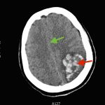
The patient’s head CT showed a significant area of hyperdensity consistent with an intracranial hemorrhage located within the left frontal parietal lobe (red arrow). Additionally, there is rightward midline shift up to 1.1cm (green arrow) and entrapment of the right lateral ventricle (blue arrow).
Discussion:
Intraparenchymal hemorrhage (IPH) is associated with high morbidity and mortality. Although the mortality for subarachnoid hemorrhage (SAH) has declined steadily over the past several decades, the mortality for IPH mortality has not significantly.1 One of the most serious considerations when treating a patient with IPH is the management of intracranial pressure (ICP).2 Once an IPH is identified, immediate steps should be taken to bring ICP within acceptable levels including elevating the head of the bed to 30 degrees, sedation, and controlling hypertension with medications.2-3 Even with early and aggressive care, the prognosis for IPH remains poor; the 30-day mortality rate for IPH is estimated to be less than 50%, and a 2010 systematic review estimated only 12-39% of IPH patients achieve independent function.4-5 Predictors of worse outcome include location of hemorrhage, level of consciousness, patient age, preceding anticoagulation therapy, and size of hematoma.5
Topics:
Intracranial hemorrhage (ICH), intraparenchymal hemorrhage (IPH), stroke, brain bleed, neurological deficits, emergency medicine.
References:
- Manno EM, Atkinson JL, Fulgham JR, Wijdicks EF. Emerging medical and surgical management strategies in the evaluation and treatment of intracerebral hemorrhage. Mayo Clin Proc. 2005;80(3):420-433. doi: 10.4065/80.3.420
- Huff JS, Wintermark M, Ghaemmaghami CA. Intracranial hemorrhages. In: Adams JG, Barton ED, Collings JL, DeBlieux PM, Gisondi MA, Nadel ES, eds. Emergency Medicine: Clinical Essentials. Philadelphia, PA: Elsevier;2013:896-904
- Broderick J, Connolly S, Feldmann E, Hanley D, Kase C, Krieger D, et al. Guidelines for the management of spontaneous intracerebral hemorrhage in adults: 2007 update: a guideline from the American Heart Association/American Stroke Association Stroke Council, High Blood Pressure Research Council, and the Quality of Care and Outcomes in Research Interdisciplinary Working Group. Stroke. 2007;38(6):2001-2023. doi: 10.1161/STROKEAHA.107.1836892
- van Asch CJ, Luitse MJ, Rinkel GJ, van der Tweel I, Algra A, Klijn CJ. Incidence, case fatality, and functional outcome of intracerebral haemorrhage over time, according to age, sex, and ethnic origin: a systematic review and meta-analysis. Lancet Neurol. 2010;9(2):167-176. doi: 10.1016/S1474-4422(09)70340-03
- Broderick JP, Brott TG, Duldner JE, Tomsick T, Huster G. Volume of intracerebral hemorrhage. A powerful and easy-to-use predictor of 30-day mortality. Stroke. 1993;24(7):987-993.


