Computed Tomography Diagnosis of Appendicitis
History of present illness:
A 19-year-old male with no previous medical history presented with 7/10 non-radiating, constant, sharp, periumbilical pain associated with nausea, and four episodes of vomiting. He was seen at urgent care where his labs showed a WBC of 16,000 x 103/mm3. He was subsequently sent to the emergency department (ED) for concern of appendicitis. Of note, his pain worsened with bumps during the drive to the ED. After arriving to the ED the pain migrated to the right lower quadrant. Computed tomography (CT) revealed acute appendicitis and the patient was admitted to the surgery service and taken to the operating room for an appendectomy.
Significant findings:
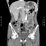
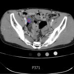
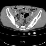
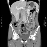
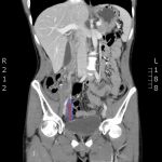
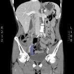
The CT abdomen/pelvis with intravenous contrast shows a dilated appendix (see red outline) with thickened, hyperenhancing wall (see blue outline) best visualized in the axial and coronal planes.
Discussion:
Appendicitis is a common diagnosis in the emergency department in patients presenting with abdominal pain, occurring most frequently in young adults with a peak incidence in those aged 10-19.1 Failure to quickly diagnose acute appendicitis can result in perforation rates as high as 80 percent.2 While the diagnosis of appendicitis can be made clinically, CT is a non-invasive modality that improves the detection of appendicitis with sensitivities of 88–100%, specificities of 91–99%, positive predictive values of 92–98%, negative predictive values of 95–100%, and accuracies of 94–98%.3-8 The major advantage of CT over both clinical exam and ultrasound is the ability of the radiologist to exclude acute appendicitis if the appendix appears normal. However, CT carries the risks associated with ionizing radiation. While previously there was some debate on the best choice for type of CT scan and use of IV and oral contrast, recent studies have shown that CT abdomen/pelvis with IV contrast alone is sufficient for diagnosis of appendicitis.9, 10
Topics:
Appendicitis, appy, CT, right lower quadrant abdominal pain, abdominal pain.
References:
- Addiss DG, Shaffer N, Fowler BS, Tauxe RV. The epidemiology of appendicitis and appendectomy in the United States. Am J Epidemiol. 1990;132(5):910-925.
- Bickell NA, Aufses AH, Rojas M, Bodian C. How time affects the risk of rupture in appendicitis. J Am Coll Surg. 2016;202:401-406. doi: 10.1016/j.jamcollsurg.2005.11.016
- Lane MJ, Katz DS, Ross BA, Clautice-Engle TL, Mindelzun RE, Jeffrey RB Jr. Unenhanced helical CT for suspected acute appendicitis. AJR Am J Roentgenol. 1997;168(2):405-409. doi: 10.2214/ajr.168.2.9016216
- Rao PM, Rhea JT, Novelline RA. Helical CT combined with contrast material administered only through the colon for imaging of suspected appendicitis. AJR Am J Roentgenol. 1997;169(5):1275-1280.
- Rao PM, Rhea JT, Noveline RA, McCabe CJ, Lawrason JN, Berger DL, et al. Helical CT technique for the diagnosis of appendicitis: prospective evaluation of focused appendix technique. Radiology. 1997;202(1):139-144.
- Walker S, Haun W, Clark J, McMillin K, Zeren F, Gilliland T. The value of limited computed tomography with rectal contrast in the diagnosis of acute appendicitis. Am J Surg. 2000;180(6):450–454.
- Ujiki MB, Murayam KM, Cribbins AJ, Angelos P, Dawes L, Prystowsky JB, et al. CT scan in the management of acute appendicitis. J Surg Res. 2002;105(2):119-122.
- Willekens I, Peeters E, De Maeseneer MD, Mey J. The normal appendix on CT: Does size matter? PLoS One. 2014;9(5): e96476. doi: 10.1371/journal.pone.0096476
- Drake FT, Alfonso R, Bhargava P, Cuevas C, Dighe MK, Florence MG, et al. Enteral contrast in the computed tomography diagnosis of appendicitis. Comparative effectiveness in a prospective surgical cohort. Ann Surg. 2014;260(2):311-316. doi: 10.1097/SLA.0000000000000272
- Servaes S, Srinivasan A, Pena A, del Pozo G, Darge K. CT diagnosis of appendicitis in children: comparison of orthogonal planes and assessment of contrast opacification of the appendix. Pediatr Emerg Care. 2015;31(3):161-163. doi: 10.1097/PEC.0000000000374

