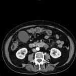
Choledocholithiasis
History of present illness:
A 75-year-old female presented with jaundice and abdominal pain for 3 weeks. She denied any fevers, confusion, or urinary complaints. On exam, she was afebrile and hemodynamically normal, but had focal tenderness in her right upper quadrant. Her neurologic exam was normal.
Significant findings:
Computed tomography (CT) was significant for two large gallstones measuring 1.1 centimeters impacted at the level of the pancreatic head with associated common bile duct (CBD) dilatation.
Discussion:
Choledocholithiasis is characterized by the presence of gallstones in the CBD. As one ages, the common bile duct enlarges, facilitating the passage of a gallstone into the CBD increasing the likelihood of developing choledocholithiasis.1 Complications associated with choledocholithiasis include pancreatitis, biliary cirrhosis, and acute cholangitis. Charcot’s triad (fever, right upper quadrant pain, and jaundice) is the classic presentation for Acute Cholangitis; however, only 50%-75% of patients have all three symptoms.2 Significant mortality is associated with Reynaud’s pentad, or the development of fever, right upper quadrant pain, jaundice, hypotension, and altered mental status.3
Sonographic measurement of CBD diameter, from inner wall to inner wall, is often sufficient for the diagnosis. Normal diameter increases 1 mm with every decade of life, with 8.5 mm having been described as the upper limit of normal.4 However, CBD dilatation up to 10 mm after cholecystectomy has been described.5 Sensitivity of an enlarged CBD in diagnosing choledocholithiasis varies from 20%-90%. 6 In some cases, CBD dilatation is not recognized on ultrasound, but is later found on CT imaging. Sensitivity of CT has been documented as high as 65%-93% with specificities approaching 100%.7
Due to lower morbidity and mortality rates, endoscopic retrograde cholangiopancreatography (ERCP) is the preferred modality for decompression of the biliary system. Some studies quote mortality rates of ERCP as low as 4.7%-10% compared to 10%-50% with open surgical decompression.8,9
Topics:
Abdominal, gastroenterology, GI, gallstone, gallbladder, choledocholithiasis, common bile duct, CBD, endoscopic retrograde cholangiopancreatography, ERCP.
References:
- Collins C, Maguire D, Ireland A, Fitzgerald F, O’Sullivan GC. A prospective study of common bile duct calculi in patients undergoing laparoscopic cholecystectomy: natural history of choledocholithiasis revisited. Ann Surg. 2004;239:28-33. doi: 10.1097/01.sla.0000103069.00170.9c
- Saik RP, Greenburg AG, Farris JM, Peskin GW. Spectrum of cholangitis. Am J Surg. 1975;130(2):143-150. doi: 10.1016/0002-9610(75)90362-1
- DenBesten L, Doty JE. Pathogenesis and management of choledocholithiasis. Surg Clin North Am. 1981;61(4):893-907.
- Bachar GN, Cohen M, Belenky A, Atar E, Gideon S. Effect of aging on the adult extrahepatic bile duct: A sonographic study. J Ultrasound Med. 2003;22(9):879-882. doi: 10.7863/jum.2003.22.9.879
- Cronan JJ. US diagnosis of choledocholithiasis: a reappraisal. Radiology. 1986;161(1):133-134. doi: 10.1148/radiology.161.1.3532178
- ASGE Standards of Practice Committee, . The role of endoscopy in the evaluation of suspected choledocholithiasis. Gastrointestinal Endosc. 2010;7(1)1:1-9. doi: 10.1016/j.gie.2009.09.041
- Tseng CW, Chen CC, Chen TS, Chang, FY, Lin HC, Lee SD. Can computed tomography with coronal reconstruction improve the diagnosis of choledocholithiasis? J Gastroenterol Hepatol. 2008;23:1586-1589. doi: 10.1111/j.1440-1746.2008.05547.x
- Lai EC, Mok FP, Tan ES, Lo CM, Fan ST, You KT, et al. Endoscopic biliary drainage for severe acute cholangitis. N Engl J Med. 1992;326(24):1582-1586. doi: 10.1056/NEJM199206113262401
- Lai EC, Tam PC, Paterson IA, Ng MM, Fan ST, Choi TK, et al. Emergency surgery for severe acute cholangitis. The high-risk patients. Ann Surg. 1990;211(1):55-59.


