Cardiology/Vascular
Endocarditis
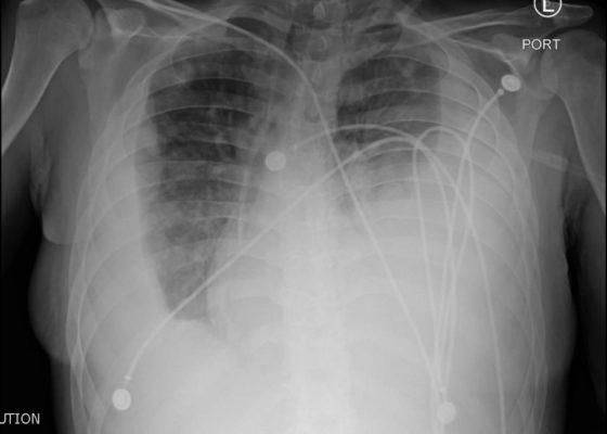
DOI: https://doi.org/10.21980/J8JP73Upright frontal radiograph of the chest demonstrated large pleural effusion on the left and moderate pleural effusion on the right as shown by the visible menisci on both sides (red arrows) with diffuse bilateral nodular densities (yellow dotted lines), consistent with septic pulmonary emboli. Computed tomography (CT) of the chest demonstrated multiple scattered lung nodules bilaterally containing internal foci of air cavitation (green dotted lines).
Clinical Evaluation and Management of Pediatric Pericarditis
DOI: https://doi.org/10.21980/J8HP85An electrocardiogram (ECG) was concerning for ST segment elevation in leads II, III, aVF, and V4, with subtle ST elevations in V5 and V6 (see black arrows). There is also ST segment depression in aVL (see blue arrows).
An Unusual Case of Hematemesis
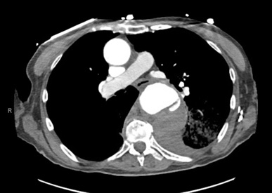
DOI: https://doi.org/10.21980/J84H00The patient’schest X-ray revealed a prominent mediastinum and opacification in the left middle and lower lung fields. The CT showed an aortic aneurysm extending from the thorax to the abdomen with rupture near T7 (blue arrow). It also showed periaortic hemorrhage with active extravasation (green arrow) likely secondary to a penetrating ulcer and bilateral pulmonary opacities concerning for hemothorax (pink arrow).
Extensive Aortic Dissection with Normal Vital Signs
DOI: https://doi.org/10.21980/J80S6SThe patient was found to have a Stanford type A dissection (see yellow arrow) with visible false lumen starting at aortic arch (see green circle). The dissection extended into the descending aorta (see blue circle) as shown by the false lumen (red highlighted area) visible on CT. The radiologist performed a reconstruction of the aorta, which showed that the left kidney was not being perfused, making the kidney not visible on the reconstruction.
Don’t Forget the Pacemaker – A Rare Complication
DOI: https://doi.org/10.21980/J8GS7HThe ECG demonstrated the presence of pacemaker spikes without appropriate capture (green arrows) and a ventricular escape rhythm which can be identified by an absence of P waves prior to the QRS complex (purple arrows). The portable chest X- demonstrated displaced pacemaker leads (red arrows) that were coiled around the pulse generator (blue arrow).
Novel Emergency Medicine Curriculum Utilizing Self-Directed Learning and the Flipped Classroom Method: Cardiovascular Emergencies Small Group Module
DOI: https://doi.org/10.21980/J8X334We aim to teach the presentation and management of cardiovascular emergencies through the creation of a flipped classroom design. This unique, innovative curriculum utilizes resources chosen by education faculty and resident learners, study questions, real-life experiences, and small group discussions in place of traditional lectures. In doing so, a goal of the curriculum is to encourage self-directed learning, improve understanding and knowledge retention, and improve the educational experience of our residents.
Right Ventricular Dilation in Patient With Submassive Pulmonary Embolism
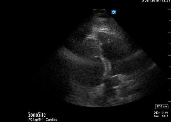
DOI: https://doi.org/10.21980/J82P84Bedside echocardiography four chamber view revealed enlarged right ventricular (RV) to left ventricular (LV) ratio (greater than 1) on apical four-chamber view (see red and blue outlines respectively). The right atrium is not clearly delineated in this image and therefore is not outlined. One can also rule out a large pericardial effusion as the cause of her dyspnea, since there is no large hypoechoic collection surrounding the heart on either four- chamber view or parasternal long view.
Fainting Spells
DOI: https://doi.org/10.21980/J8Z91RABSTRACT: Audience: The target audience for this simulation is 4th year medical students, emergency medicine residents, pediatric residents, and family medicine residents. Introduction: Brugada syndrome is defined as the combination of specific electrocardiogram (ECG) changes and clinical manifestations of a ventricular arrhythmia, including syncope and sudden cardiac arrest.1 Brugada syndrome is caused by a mutation in the phase-0 cardiac sodium channel. This


