
Cardiology/Vascular
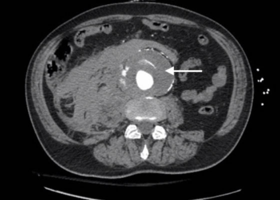

Ruptured AAA Presenting with Unresponsiveness and Cardiac Arrest
DOI: https://doi.org/10.21980/J8M34QAxial CT images from the CT chest, abdomen and pelvis revealed a large infra-renal abdominal aortic aneurysm measuring 7.3 x 8.2 x 10 cm with extensive mural thrombus (single white arrow) that has ruptured, with active extravasation (black arrow) of contrast into the aneurysm sac and retroperitoneum with large right retroperitoneal hematoma (multiple white arrows).

Levamisole Induced, Cocaine Associated Vasculitis
DOI: https://doi.org/10.21980/J8K35SAn asymmetric pattern of palpable purpura with bullae was noted on bilateral lower extremities with smaller patches on bilateral upper extremities. There was no tenderness or crepitus.
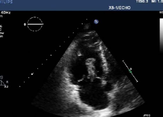
Right Atrial Thrombus
DOI: https://doi.org/10.21980/J8F93V History of present illness: A 77-year-old male presented to the emergency department with shortness of breath. Symptoms progressively worsened over the last 4-5 days, and on arrival was associated with chest tightness. He denied any medical conditions, smoking, or pertinent family history. He has not seen a primary care physician in “many years.” Upon arrival he was in mild

Point-of-Care Ultrasound for the Diagnosis of Systolic Heart Failure
DOI: https://doi.org/10.21980/J8HD1RBedside ultrasound with the phased array probe was used to obtain a parasternal long axis view which demonstrated poor contractility and a severely decreased ejection fraction (EF). M-mode was placed over the anterior leaflet of the mitral valve to create a tracing depicting both the E-wave of early diastole (green arrow) and the A-wave from the atrial kick (blue arrow). The shortest distance between the septum and the mitral valve on the M-mode tracing gives the patient’s E-Point Septal Separation (EPSS) (pink arrow). EF can be estimated using the formula EF=75.5-2.5 x EPSS (in mm). This patient’s EPSS was measured to be 20mm which estimates that she had an EF of 25.5%.
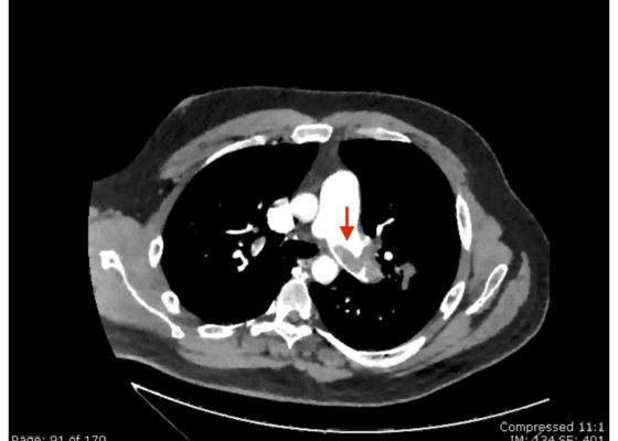
Saddle Pulmonary Embolus
DOI: https://doi.org/10.21980/J8N63PAn electrocardiogram (ECG) showed evidence of right heart strain with an incomplete right bundle branch block, S1Q3T3 (see red arrow [S1], blue arrow [Q3], and black arrow [T3]), and ST-segment elevation in the septal leads (green arrows). Bedside echocardiography showed a dilated right ventricle with ventricular wall akinesis (red arrow) sparing the apex (purple arrow), which is known as McConnell’s Sign. It also showed a mobile hyperechoic mass (yellow arrow). These ultrasound findings were concerning for pulmonary embolism (PE), so computed tomography (CT) angiogram of the chest was ordered and confirmed massive bilateral obstructive filling defects (red arrows) consistent with saddle pulmonary embolism. Additionally, noted is flattening of the interventricular septum (blue arrow) consistent with right heart strain. Laboratory studies were notable for a troponin-I of 0.29 ng/mL, a B-type natriuretic peptide of 792.3 pg/mL, lactic acid of 5.30 mmol/L, and a creatinine of 2.0 mg/dL, consistent with end organ dysfunction. All other lab work was within normal limits.
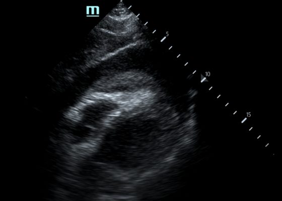
Pericardial Clot on Point-of-Care Ultrasound
DOI: https://doi.org/10.21980/J8ZH1TFocused assessment with sonography in trauma (FAST) scan was positive for a clinically significant pericardial effusion as evidenced by the hypoechoic fluid around the myocardium, indicated by the blue arrow in image 2. Findings are also consistent with tamponade process as evidenced by restricted expansion and collapse of the right ventricle during diastole. The hyperechoic floating structure between the pericardium and myocardium, adjacent to the right ventricle, represents a pericardial clot, indicated by the white arrow.The density of the pericardial clot differs from that of the myocardium, thus serving as an additional variable to avoid confusing this as part of the myocardial structure.

Wellens’ Syndrome
DOI: https://doi.org/10.21980/J8FS8KInitial electrocardiogram (ECG) revealed the classic biphasic T waves in V2 and V3 of Wellen’s syndrome (see red outlines). A second EKG demonstrated an evolving deeply inverted T wave (see blue outlines).

Arteriovenous Graft Pseudoaneurysm
DOI: https://doi.org/10.21980/J8B06ZA bedside ultrasound of the mass demonstrated a large compressible hypoechoic structure (see purple outline) above the arteriovenous graft (see red outline). The contents demonstrated movement of fluid within the structure. This was confirmed with Doppler mode, which allowed for visualization of flow communicating between the structure and the underlying vessel, which is diagnostic for a pseudoaneurysm.