Idiopathic Intracranial Hypertension and Optic Nerve Sheath Diameter
History of present illness:
A 38-year-old female with body mass index (BMI) of 47.3 and history of insulin-dependent diabetes mellitus presented to the emergency department with headache. She described the headache as slowly progressive over three weeks with “pressure-like” quality and 9/10 severity at the back of her head with associated blurred vision. Physical exam revealed normal vital signs, normal neurologic exam, intact visual fields, no nuchal rigidity, and a normal fundoscopic exam except for mild papilledema bilaterally.
Significant findings:
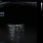
Optic nerve sheath diameter (ONSD) was measured via ultrasound with diameter 5.7mm on left and 6.2mm on right. In order to measure ONSD via optic ultrasound the high-frequency linear array probe (7.5-10-MHz or higher) is utilized in B-mode. The patient is positioned supine and an occlusive dressing, such as Tegaderm, is placed over a closed eyelid with copious conductive gel on top of the dressing. Being careful not to put pressure on the globe, an axial cross-sectional image of the globe is obtained. As demonstrated in the image “annotated left eye ONSD pre-lumbar puncture,” there are two main anechoic areas of the globe, the anterior chamber and the vitreous humor. These anechoic structures are separated by the hyperechoic iris, which surfaces the hyper-echoic-lined lens. At the back of the vitreous humor is the retina, which leads posteriorly into the optic nerve. The optic nerve is the hypoechoic structure posterior to the retina and surrounded by the hyperechoic subarachnoid space, which is encased by the hypoechoic dura mater. The outer edge of the hypoechoic dura matter is where the ONSD is measured.1 The user applies calipers to measure 3mm perpendicularly behind the retina along the hypoechoic optic nerve, and at this level the transverse dimensions of the ONSD are measured using calipers as shown in the images.
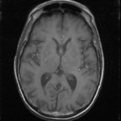
Computed tomography (CT) of the head was performed and showed no abnormalities. Lumbar puncture was performed in left lateral decubitus position revealing elevated opening pressure of 29cm H2O. Thirty-five mL of clear cerebral spinal fluid was drained and was negative for all infectious studies. Optic nerve sheath diameter was again measured post-lumbar puncture with diameters 5.4mm on left and 5.4mm on right.
Discussion:
Idiopathic intracranial hypertension (IIH) is a vision-threatening disease characterized by signs of increased intracranial pressure including headache and papilledema without any secondary cause.2 The diagnostic criterion for IIH is outlined by the modified Dandy Criteria (Table 1).3 Ultrasonography of the optic nerve sheath diameteris a non-invasive way to monitor intracranial pressure (ICP), and its accuracy, in comparison to gold standard measurement via invasive intracranial devices in the ventricles or cerebral parenchyma, has been shown in a handful of studies. The rational for using ONSD as a surrogate for invasive intracranial monitoring is as follows: increased ICP is transmitted to the subarachnoid space surrounding the optic nerve; this causes the sheath around the nerve to expand with cerebrospinal fluid, and this expansion may be measured directly via ultrasound.1
In comparing ONSD to invasive gold standard intraventricular or intraparenchymal ICP monitoring in traumatic brain injury or hemorrhage patients, a 2011 meta-analysis found sensitivity 90% and specificity 85% of ONSD compared to invasive monitoring.4 A systematic review and meta-analysis by Robba, et al, compared ultrasound to CT in identifying elevated ICP and found that ultrasound was 95.6% sensitive and 92.3% specific with a 0.05% negative likelihood ratio compared to CT.5 Previous studies have identified ONSD greater than 5mm as the cut-off point suggestive of elevated ICP.6 A prospective observational single-center study examined neurosurgical intensive care patients who had invasive intracranial pressure monitoring in place via intraparenchymal catheter or external ventricular drain catheters and compared these measurements to those found via ultrasonographic ONSD.7Among both traumatic and non-traumatic subjects, this study found that ONSD of 5.2mm was 95.8% sensitive and 80.4% specific for raised ICP. However, when the study was divided to only include non-traumatic patients, patients who are more representative of an IIH patient, a cut off of 5.2mm yielded increased sensitivity at 100%, but decreased specificity at 76.7%. Although this study compared ONSD directly to the gold standard measurement for ICP, this study is limited in generalizability because it did not include IIH patients. A recent single-center case control study from 2018 evaluated ONSD in IIH patients and healthy controls and determined a cut-off value of 6.05mm for IIH, which yielded sensitivity 73.2% and specificity 91.4%.1 In this study, cut-off for 100% sensitivity was 3.1mm and 100% specificity was 6.5mm.
These studies demonstrate a likely correlation between elevated ICP as measured by ONSD in IIH patients; however, to safely determine a cut-off value for ONSD above which IIH should be suspected and to determine if ONSD may be used to monitor IIH treatment following lumbar puncture in the emergency department, more studies are needed. For now, this should not change our practice, but this may be used as an adjunct to our typical emergency department evaluation of IIH, such as fundoscopic exam, CT, and lumbar puncture.
For this patient, a CT was performed as well as ONSD measurements before and after the diagnostic and therapeutic lumbar puncture. Following lumbar puncture, the measurements of the patient’s ONSD decreased and the patient’s symptoms improved. She was discharged on acetazolamide and with an outpatient neurologic follow-up appointment. Medical treatment for IIH includes weight loss and acetazolamide.1 Further interventions by neurology and neurosurgery for medically refractory cases include serial lumbar punctures, optic nerve sheath fenestration, or CSF shunting.8 However, for either acute or medically refractory cases of possible IIH presenting to the emergency department, such as in this case, diagnosis and treatment is via normal brain imaging and lumbar puncture with elevated opening pressure, negative CSF studies, and symptom relief.9 The therapeutic removal of CSF via lumbar puncture aims to relieve elevated ICP symptoms while avoiding post lumbar puncture headaches. To achieve this, it is suggested to aim for a high normal closing pressure of approximately 18-20cm H2O and to do so by draining 1mL CSF for every 1.5cm H2O of desired opening pressure decrease.10 The utility of ONSD as an additional way to detect IIH and monitor efficacy of medical and interventional treatment seems promising, but requires further investigation.
Topics:
Neurology, intracranial, idiopathic intracranial hypertension, ultrasound, optic nerve, optic nerve sheath diameter.
References:
- Kishk NA, Ebraheim AM, Ashour AS, Badr NM, Eshra MA. Optic nerve sonographic examination to predict raised intracranial pressure in idiopathic intracranial hypertension: The cut-off points. Neuroradiol J.2018;31(5):490-495. doi: 10.1177/1971400918789385.
- Wall M, Kupersmith MJ, Kieburtz KD, et al. The idiopathic intracranial hypertension treatment trial: clinical profile at baseline. JAMA Neurol. 2014;71(6):693-701. doi: 10.1001/jamaneurol.2014.133.
- Friedman DI, Jacobson DM. Diagnostic criteria for idiopathic intracranial hypertension. Neurology. 2002;59(10):1492-1495. doi: 10.1212/01.WNL.0000029570.69134.1B.
- Dubourg J, Javouhey E, Geeraerts T, Messerer M, Kassai B. Ultrasonography of optic nerve sheath diameter for detection of raised intracranial pressure: a systematic review and meta-analysis. Intensive Care Med. 2011;37(7):1059-1068. doi: 10.1007/s00134-011-2224-2.
- Robba C, Santori G, Czosnyka M, et al. Optic nerve sheath diameter measured sonographically as non-invasive estimator of intracranial pressure: a systematic review and meta-analysis. Intensive Care Med. 2018;44(8):1284-1294. doi: 10.1007/s00134-018-5305-7.
- Kimberly HH, Shah S, Marill K, Noble V. Correlation of optic nerve sheath diameter with direct measurement of intracranial pressure. Acad Emerg Med. 2008;15(2):201-4. doi: 10.1111/j.1553-2712.2007.00031.x.
- Raffiz M, Abdullah JM. Optic nerve sheath diameter measurement: a means of detecting raised ICP in adult traumatic and non-traumatic neurosurgical patients. Am J Emerg Med. 2017;35(1):150-153. doi: 10.1016/j.ajem.2016.09.044.
- Piper RJ, Kalyvas AV, Young AM, Hughes MA, Jamjoom AA, Fouyas IP. Interventions for idiopathic intracranial hypertension. Cochrane Database Syst Rev. 2015;(8):CD003434. doi: 10.1002/14651858.CD003434.pub3.
- Bidot S, Bruce BB. Update on the Diagnosis and Treatment of Idiopathic Intracranial Hypertension. Semin Neurol. 2015;35(5):527-38. doi: 10.1055/s-0035-1563569.
- Parikh S, Fiorito-Torres F, Rayhill M, McAdams M, Perloff M.Updated Results on Cerebrospinal Fluid (CSF) Volume Removal for Idiopathic Intracranial Hypertension (IIH): Removing Less CSF May Be Best (P3.101). Neurology. Apr 2018, 90(15);101.


