Issue 6:3
A Case Report of a Large Goiter Resulting in Tracheal Deviation
DOI: https://doi.org/10.21980/J80645In the image, one can see significant tracheal deviation around the right side of the mass (black arrows). This degree of deviation would make ventilation in a paralyzed patient extremely difficult, if not impossible.
Case Report—Pediatric Brugada Phenotype from Accident Cocaine Ingestion
DOI: https://doi.org/10.21980/J8VH28Initial EKG was concerning for type I Brugada pattern with an incomplete right bundle branch block in V1 & ST segment elevation terminating in an inverted T wave in V2. There are also signs of sodium channel toxicity with a widened QRS complex, tachycardia and a terminal R wave present in aVR where the R wave is bigger than the S wave or the R wave is over 3mm in aVR.
A Case Report of Neonatal Vomiting due to Adrenal Hemorrhage, Abscess and Pseudohypoaldosteronism
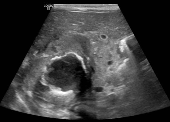
DOI: https://doi.org/10.21980/J8QQ0BAn ultrasound (US) of the abdomen was obtained to evaluate for possible pyloric stenosis (see US transverse/dopper imaging). While imaging showed a normal pyloric channel, it revealed an unexpected finding: a complex cystic mass arising from the right adrenal gland (yellow outline), measuring 5.8 by 4.0 by 6.4 cm with calcifications peripherally and mass effect on the kidney without evidence of vascular flow (blue arrow). Computed tomography (CT) of the abdomen/pelvis with IV contrast was subsequently obtained and measured the lesion as 2.8 by 4.6 by 4 cm without evidence of additional masses, lymphadenopathy or left adrenal gland abnormality (see CT axial, coronal, and sagittal imaging).
Case Report of Unusual Facial Swelling in an 8-Month-Old
DOI: https://doi.org/10.21980/J8M06FFacial ultrasound revealed local inflammatory changes such as increased echogenicity and heterogeneity in the soft tissues of the right cheek, suggestive of soft tissue edema. There was evidence of a prominent right parotid gland with increased heterogeneity suggestive of a traumatic injury. Additionally, facial ultrasound demonstrated a 6mm ill-defined anechoic collection within the right cheek without increased doppler flow (green arrow), thought to represent a focal area of edema instead of an abscess.
Case Report: Thoracic Aortic Dissection in a Previously Healthy Male with an Unusual Inciting Factor
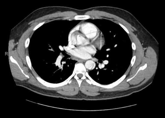
DOI: https://doi.org/10.21980/J8G92SA non-contrast computed tomography (CT) scan was negative for a suspected ureteral stone. However, there were aortic calcifications visualized at the infrarenal level that were notable given the patient’s age (red arrow). Given this finding in conjunction with the patient’s symptoms, bedside transabdominal ultrasonography was performed which revealed an intraluminal echogenic flap within the aorta near the common iliac arteries. CT angiography (CTA) with delayed contrast protocol revealed an extensive Stanford type A aortic dissection with involvement of the aortic root (purple arrow), brachiocephalic trunk, ostia of the left subclavian artery, descending aorta (blue arrow), bilateral common iliac arteries, and left internal iliac artery.
‹2
Page 2 of 2


