Caught on CT! The Case of the Hemodynamically Stable Ruptured Abdominal Aortic Aneurysm
ABSTRACT:
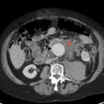
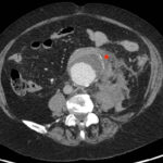
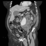
Rupture of an abdominal aortic aneurysm (AAA) is a surgical emergency with significant associated mortality. This case report discusses a 70-year-old woman presenting to the emergency department (ED) with vague and progressive abdominal pain and relatively benign vital signs and physical exam. She was ultimately diagnosed with a ruptured infrarenal 6.8cm abdominal aortic aneurysm seen on computerized tomography (CT) imaging. Found to be stable, she was transferred to an affiliate tertiary care facility and underwent endovascular repair of the aneurysm. The surgery was successful, and she has since recovered nicely. As reported at her most recent follow up appointments. A timely diagnosis of a ruptured AAA is vital for any chance of a successful outcome. However, in patients with normal vital signs and an unremarkable physical exam, this diagnosis can be incredibly difficult. As such, the need for a broad differential in the setting of abdominal pain is imperative to avoid missing the diagnosis. The use of point of care ultrasound (POCUS) can further aid in this process by providing a quick and affordable screening option to rule out potential aneurysms.
Topics:
Abdominal Aortic Aneurysm, Ruptured Abdominal Aortic Aneurysm, CT imaging, Tangential Calcium Sign, Point of Care Ultrasound.


