Urology
Difficult Conversation Case: Missed Testicular Cancer
DOI: https://doi.org/10.21980/J8.52336This difficult conversation case is intended to assess the examinee’s ability to disclose sensitive, unexpected information to a patient regarding a missed diagnosis of testicular cancer. By the end of this session, learners should be able to, 1) demonstrate effective communication, including establishing rapport, acknowledging a prior misdiagnosis, and disclosing a revised diagnosis of cancer, 2) elicit and react to the patient’s emotional and informational needs in an empathetic and professional manner, and 3) convey a patient-centered plan of care, including appropriate next steps and coordination with specialist services.
The EMazing Race: A Novel Gamified Board and Clinical Practice Review for Emergency Medicine Residents
DOI: https://doi.org/10.21980/J8.52075By the end of this 2-hour session, learners will demonstrate their knowledge on the following board-related emergency medicine topics: Ob/GYN – links to 13.7 Complications of Delivery in Core Model of EM 2022, Renal/GU – links to 15.0 Renal and Urogenital Disorders in Core Model of EM 2022 and Splinting – links to 18.1.8.2 Extremity bony trauma, fracture in Core Model of EM 2022.
A Case Report of Fournier’s Gangrene
DOI: https://doi.org/10.21980/J8Z356Physical exam revealed a comfortable-appearing male patient with tachycardia and a regular cardiac rhythm. The genitourinary exam indicated significant erythema and fluctuance of the bilateral lower buttocks with extension to the perineum. Black eschar and ecchymosis were also noted at the perineum. There was significant tenderness to palpation that extended beyond the borders of erythema. There was no palpable crepitus on initial examination. Physical exam was otherwise unremarkable.
The Zipperator! A Novel Model to Simulate Penile Zipper Entrapment
DOI: https://doi.org/10.21980/J8NS8FAfter training on the Zipperator, learners will be able to: 1) demonstrate at least two techniques for zipper release and describe how methods would extrapolate to a real patient; 2) verbalize increased comfort with the diagnosis of zipper entrapment; and 3) present a plan of care for this low-volume, high-anxiety presentation.
An Atraumatic, Idiopathic Case Report of Intraperitoneal Bladder Dome Rupture
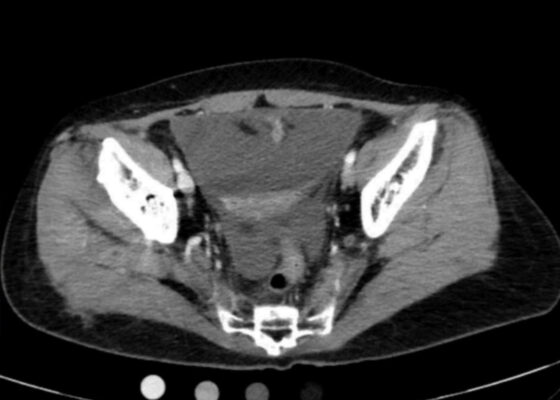
DOI: https://doi.org/10.21980/J85S83On regular CT scan imaging, the urinary bladder is partially distended with contrast with no focal wall thickening or intraluminal hematoma. There is an intraperitoneal bladder rupture with site of rupture likely at the dome of the bladder. The bladder is outlined in red, and the bladder rupture boundaries are outlined in yellow, showing the urine as free fluid escaping into the intraperitoneal space. We also provide these findings in an axial CT in video format. On CT cystography, there is a significant amount of contrast-enhanced urine noted within the visualized peritoneal spaces. The small amount of air present anteriorly is related to the catheterization because a Foley balloon is present within the bladder. These findings are annotated with the peritoneal spaces outlined in yellow, the air in the blue outline, and the bladder in the red outline. All of these CT cystography findings are also presented in an axial view in video format.
Ureteral Obstruction and Ureteral Jet Identification—A Case Report
DOI: https://doi.org/10.21980/J8206GA point-of-care ultrasound of the urinary tract was performed, evaluating the kidneys and bladder. When imaging her kidneys, right-sided hydronephrosis was noted with a normal appearance to the left kidney. To further evaluate, a curvilinear probe was placed on her bladder with color doppler to assess for ureteral jets. Ureteral jets are seen as a flurry of color ejecting from each of the ureters as urine is released from the ureterovesical junction. In a healthy patient, this finding should be seen ejecting from both ureters every 1-3 minutes as the kidneys continue to filter the blood and create urine to be stored in the bladder. In our patient, however, ureteral jets were only noted on the left side (arrow), which was significant in further verifying our suspicion of right ureteral obstruction.
Design and Implementation of a Low-Cost Priapism Reduction Task Trainer
DOI: https://doi.org/10.21980/J8K64FBy the end of this educational session, learners should be able to 1) Verbalize the difference between low-flow and high-flow priapism 2) Describe the landmarks for a penile ring block and cavernosal aspiration/injection 3) Demonstrate the appropriate technique for performing a penile ring block, cavernosal aspiration, and cavernosal injection.


