• Registration
  • Login
JETem
  • Home
  • About
    • Aim and Scope
    • Our Team
    • Editorial Board
    • FAQ
  • Issues
    • Current Issue
    • Ahead of Print
    • Past Issues
  • Visual EM
    • Latest Visual EM
    • Search Visual EM
    • Thumbnail Library
  • For Authors
    • Instructions for Authors
    • Submit to JETem
    • Photo Consent
    • Policies
      • Peer Review Policy
      • Copyright Policy
      • Editorial Policy, Ethics and Responsibilities
      • Conflicts of Interest & Informed Consent
      • Open Access Policy
  • For Reviewers
    • Instructions for JETem Reviewers
    • Interested in Being a JETem Reviewer?
  • Topic
    • Abdominal / Gastroenterology
    • Administration
    • Board Review
    • Cardiology / Vascular
    • Clinical Informatics, Telehealth and Technology
    • Dermatology
    • EMS
    • Endocrine
    • ENT
    • Faculty Development
    • Genitourinary
    • Geriatrics
    • Hematology / Oncology
    • Infectious Disease
    • Miscellaneous
    • Neurology
    • Ob / Gyn
    • Ophthalmology
    • Orthopedics
    • Pediatrics
    • Pharmacology
    • Procedures
    • Psychiatry
    • Renal / Electrolytes
    • Respiratory
    • Social Determinants of Health
    • Toxicology
    • Trauma
    • Ultrasound
    • Urology
    • Wellness
    • Wilderness
  • Modality
    • Curricula
    • Innovations
    • Lectures
    • Oral Boards
      • Structured Interview
      • Communication Case
    • Podcasts
    • Simulation
    • Small Group Learning
    • Team Based Learning
    • Visual EM
  • Contact Us

Bullous Emphysema

Valerie Lew, MD*, John Costumbrado, MD, MPH^ and Steven Kim, MD^

*Washington University in St. Louis/Barnes-Jewish Hospital, Department of Emergency Medicine, St. Louis, MO

^University of California Riverside, Department of Emergency Medicine, Riverside, CA

Correspondence should be addressed to Valerie Lew, BS at valerielew6@gmail.com 

DOI: https://doi.org/10.21980/J8W62GIssue 3:3
RespiratoryVisual EM
No ratings yet.
Creative Commons images
prev
next

History of present illness:

27-year-old male presented with left chest pain and shortness of breath for 1 month. He reported sharp pain and urge to cough upon taking a deep breath. His lungs were clear bilaterally, without rhonchi, wheezing, rubs or crackles. He denied fevers and never smoked. He had an albuterol inhaler as needed for allergic wheezing.

Significant findings:

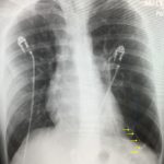
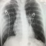
The upright chest X-ray shows a large lucent area in the left lower lung field without lung markings, with associated curvilinear opacities (yellow arrows) consistent with a large air-filled bulla. The bulla is large enough to compress adjacent lung tissue as shown by the visible pleural line (blue line). The discontinuity of the pleural line and presence of lung markings superiorly makes these findings more consistent with bulla than pneumothorax. The chest computed tomography (CT) confirmed a large left hemithorax bulla.

Discussion:

A bulla is a greater than 1 cm distended space of air in the lung.1 If the space occupies greater than 30% of the hemithorax, it is considered a giant bulla.2 The most common causes of bulla formation are smoking, chronic obstructive pulmonary disease (COPD)or idiopathic.1 Damage to the alveoli causing distension may result in the formation of larger air spaces, known as a bulla. Giant bulla are rare occurrences; thus, the epidemiology is not well characterized.3 Patients can present asymptomatically or with obstructive pulmonary symptoms such as productive cough and dyspnea. Chest X-ray is generally sufficient to diagnose a bulla, but may be mistaken as a pneumothorax, which can lead to incorrectly placing a chest tube.3

However, this is not always true, as seen in the chest X-ray above. Mediastinal structures can be deviated to the contralateral side in both a bulla or pneumothorax.4 Thus, any questionable bulla should be confirmed with a chest CT. While comparisons of imaging modalities specifically for giant bullous disease has not been well documented, there is evidence that CT is more sensitive than chest X-ray in detecting emphysematous pulmonary disease.5

For symptomatic patients, treatment of giant bulla is initially managed medically. Surgical bullectomy is considered in refractory and symptomatic idiopathic cases.6 Our patient was treated in the emergency department with ipratropium and albuterol. With treatment, his oxygen saturation improved to greater than 95% on room air. The bulla was discussed with pulmonology, who agreed this could be treated as an outpatient. The patient was discharged home with pulmonology follow-up.

Topics:

Bullous emphysema, chest radiograph, pneumothorax, bulla.

References:

  1. Thurlbeck WM. Pathophysiology of chronic obstructive pulmonary disease. Clin Chest Med. 1990;11(3):389-403.
  2. Neviere R, Catto M, Bautin N, et al. Longitudinal changes in hyperinflation parameters and exercise capacity after giant bullous emphysema surgery. J Thorac Cardiovasc Surg. 2006;132(5):1203-1207.doi: 10.1016/j.jtcvs.2006.08.002
  3. Bourgouin P, Cousineau G, Lemire P, Hébert G. Computed tomography used to exclude pneumothorax in bullous lung disease. J Can Assoc Radiol. 1985;36(4):341-342.
  4. Chen CK, Cheng HK, Chang WH, Lai YC, Su YJ. Giant emphysematous bulla mimicking tension pneumothorax. Am J Med Sci. 2009;337(3):205.doi: 10.1097/MAJ.0b013e31818713f0
  5. Uppaluri R, Mitsa T, Sonka M, Hoffman EA, McLennan G. Quantification of pulmonary emphysema from lung computed tomography images. Am J Respir Crit Care Med. 1997;156(1):248-254.
  6. Palla A, Desideri M, Rossi G, et al. Elective surgery for giant bullous emphysema: a 5-year clinical and functional follow-up. 2005;128(4):2043-2050. doi: 10.1378/chest.128.4.2043
Icon

Bullous Emphysema - Case Report

1 file(s) 606 KB
Download
Icon

Bullous Emphysema - Images

1 file(s) 6.1 MB
Download
Issue 3:3

Reviews:

No ratings yet.

Please rate this


Creative Commons images

The Role of Chest X-Ray and Bedside Ultrasound in...

14 Jul, 18
Creative Commons images

Bedside Ultrasound of Retinal Detachment in a...

14 Jul, 18

JETem is an online, open access, peer-reviewed, journal-repository for EM educators. We are PMC Indexed.

Most Viewed

  • The Silent Saboteur: Teaching the Clinical Implications of Occult Hypoxemia & Social Determinants of Health via a Pulmonary Embolism Case
  • Diabetic Ketoacidosis and Necrotizing Soft Tissue Infection
  • My Broken Heart
  • Stabilization of Cardiogenic Shock for Critical Care Transport, a Simulation
  • Innovative Ultrasound-Guided Erector Spinae Plane Nerve Block Model for Training Emergency Medicine Physicians

Visit Our Collaborators

Creative Commons Licence
This work is licensed under a Creative Commons Attribution 4.0 International License.

About

Education

Learners should benefit from active learning. JETem accepts submissions of team-based learning, small group learning, simulation, podcasts, lectures, innovations, curricula, question sets, and visualEM.

Scholarship

We believe educators should advance through the scholarship of their educational work. JETem gives educators the opportunity to publish scholarly academic work so that it may be widely distributed, thereby increasing the significance of their results.

Links

  • Home
  • Aim and Scope
  • Current Issue
  • For Reviewers
  • Instructions for Authors
  • Contact Us

Newsletter

Sign up to receive updates from JETem regarding newly published issues and findings.

Copyright Creative Commons Attribution 4.0 International

We use cookies to ensure that we give you the best experience on our website. If you continue to use this site we will assume that you are happy with it.Ok