Issue 6:4
Ureteral Obstruction and Ureteral Jet Identification—A Case Report
DOI: https://doi.org/10.21980/J8206GA point-of-care ultrasound of the urinary tract was performed, evaluating the kidneys and bladder. When imaging her kidneys, right-sided hydronephrosis was noted with a normal appearance to the left kidney. To further evaluate, a curvilinear probe was placed on her bladder with color doppler to assess for ureteral jets. Ureteral jets are seen as a flurry of color ejecting from each of the ureters as urine is released from the ureterovesical junction. In a healthy patient, this finding should be seen ejecting from both ureters every 1-3 minutes as the kidneys continue to filter the blood and create urine to be stored in the bladder. In our patient, however, ureteral jets were only noted on the left side (arrow), which was significant in further verifying our suspicion of right ureteral obstruction.
A Culinary Misadventure: A Case Report of Shiitake Dermatitis
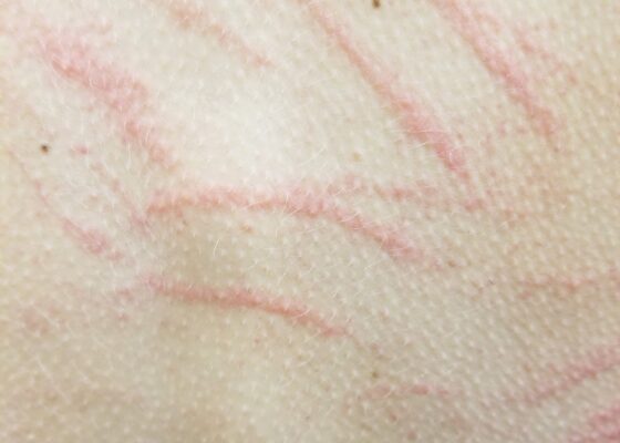
DOI: https://doi.org/10.21980/J8X936Close visual examination revealed erythematous linear papules on her upper and lower back. No bullae, drainage, or sloughing of the skin was present. The rest of her body, including palms, soles, and mucosa, was spared.
Case Report of an Empyema Identified on Lung Ultrasound
DOI: https://doi.org/10.21980/J8SH2NUsing a curvilinear ultrasound probe, images of the patient were obtained from the left mix-axillary line. These images demonstrate a loculated left-sided pleural effusion (outlined in the attached ultrasound image in blue) that was moderate in size, concerning for an empyema. The diaphragm on the right (red) of the image outlines the inferior margin of the collection of pus, which is seen in the inferior aspect of the left lung. Unfortunately, rib shadows on the left side of the image prevent the full empyema from being captured in this single image. As a result of the bedside ultrasound, however, the patient was rapidly diagnosed with an empyema and was initiated on antibiotics, which is further discussed below. After his bedside ultrasound was completed, his chest x-ray revealed the expected left-sided pleural effusion. Additionally, a CT angiogram of the chest was ordered to rule out a pulmonary embolism, which was negative for an embolism but does redemonstrate the left-sided loculated pleural effusion (outlined on the CT axial and coronal images in blue).
‹2
Page 2 of 2


