EKG
ST Elevation in aVR with Coexistent Multilead ST Depression
DOI: https://doi.org/10.21980/J8KS3XThe ECG shows ST-segment depressions in precordial leads V3 through V6, and limb leads I, II, and aVL, and 1 mm of ST-segment elevation in aVR. The initial troponin I was elevated at 1.37 ng/mL (upper limit of normal 0.40). Cardiology decided to delay catheterization until the next day when diffuse coronary disease was discovered (including 90% of the left circumflex stenosis, 60% proximal and 75% mid-left anterior descending stenosis, 75% third diagonal branch stenosis, and 90% posterior descending artery stenosis). The following day, the patient went to the operating room for coronary artery bypass grafting (CABG).
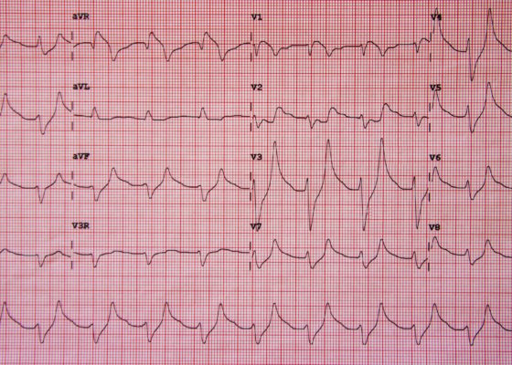
Hyperkalemia on ECG
DOI: https://doi.org/10.21980/J8K017Initial ECG shows tall, peaked T waves, most prominently in V3 and V4, as well as QRS widening. These findings are consistent with hyperkalemia, which was promptly treated. Follow-up ECG post-treatment shows narrowing of the QRS complexes and normalization of peaked T waves.


