
JETem
  • Home
  • About
    • Aim and Scope
    • Our Team
    • Editorial Board
    • FAQ
  • Issues
    • Current Issue
    • Ahead of Print
    • Past Issues
  • Visual EM
    • Latest Visual EM
    • Search Visual EM
    • Thumbnail Library
  • For Authors
    • Instructions for Authors
    • Submit to JETem
    • Photo Consent
    • Policies
      • Peer Review Policy
      • Copyright Policy
      • Editorial Policy, Ethics and Responsibilities
      • Conflicts of Interest & Informed Consent
      • Open Access Policy
      • Acceptance Fee
  • For Reviewers
    • Instructions for JETem Reviewers
    • Interested in Being a JETem Reviewer?
  • Topic
    • Abdominal/Gastroenterology
    • Administration
    • Board Review
    • Cardiology/Vascular
    • Certifying Exam Cases
      • Clinical Decision-Making
      • Prioritization
      • Communication
      • Procedure & Ultrasound
    • Clinical Informatics, Telehealth and Technology
    • Dermatology
    • EMS
    • Endocrine
    • ENT
    • Faculty Development
    • Genitourinary
    • Geriatrics
    • Hematology/Oncology
    • Infectious Disease
    • Miscellaneous (stats, etc)
    • Neurology
    • Ob/Gyn
    • Ophthalmology
    • Orthopedics
    • Pediatrics
    • Pharmacology
    • Procedures
    • Psychiatry
    • Renal/Electrolytes/Nutrition
    • Respiratory
    • Social Determinants of Health
    • Toxicology
    • Trauma
    • Ultrasound
    • Urology
    • Wellness
    • Wilderness
  • Modality
    • Certifying Exam Practice Cases
      • Clinical Care Cases
      • Communication & Procedural Cases
      • Old School Oral Board Cases
    • Curricula
    • Innovations
    • Lectures
    • Podcasts
    • Simulation
    • Small Group Learning
    • Team Based Learning (TBL)
    • Visual EM
  • Contact Us

Cardiology/Vascular

Pulseless Electrical Activity Cardiac Arrest

Erik Sembroski, MD*,  Christopher M McDowell, MD^ and Matthew M Mannion, BA^

DOI: https://doi.org/10.21980/J8Z055 Issue 5:1[mrp_rating_result]
After competing this simulation-based session, the learner will be able to: 1) Identify PEA arrest; 2) review the ACLS commonly recognized PEA arrest etiologies via the H &T mnemonic; 3) review and discuss the risks and benefits of tissue plasminogen activator (tPA) for massive PE.
Cardiology/VascularRespiratorySimulation
Creative Commons images

Bilateral Common Iliac Artery Aneurysm

Laura Kolster, DO*, Danielle Biggs, MD*, Amy Patwa, DO* and Michael Gerardi, MD*

DOI: https://doi.org/10.21980/J83S73 Issue 5:1[mrp_rating_result]
A bedside ultrasound of the aorta was performed. The proximal, middle, and distal aorta appeared normal in caliber, as demonstrated by the images; however there seemed to be some enlargement at the bifurcation. The bifurcation into the iliac arteries, as highlighted by the yellow arrow, demonstrates a slightly enlarged iliac artery on the left. The aorta was followed below the bifurcation as it divided into the iliac arteries, as shown in the video clip. The ultrasound demonstrated a left iliac artery aneurysm measuring 5.99 cm, as highlighted by the orange circle. There were aneurysms to the bilateral common and internal iliac arteries.
Cardiology/VascularAbdominal/GastroenterologyVisual EM
Creative Commons images

A Low-Cost, Reusable Ultrasound Pericardiocentesis Simulation Model

Jonathan dela Cruz, MD*, Tyler Fulks, MD*, Mark Baker, MD*, Jason Kegg, MD*, Richard Austin, MD*, Jimmy Jain, MD*, Michael Boehler, MS*, Sharon Kim, PhD^, and Cassie Jaeger, PhD^

DOI: https://doi.org/10.21980/J8TD1J Issue 4:4[mrp_rating_result]
Through the use of this model and skill session, learners will be able to: 1) discuss the indications, contraindications, and complications associated with ultrasound guided pericardiocentesis; 2) demonstrate an ability to obtain subxiphoid and parasternal long views of the heart; 3) demonstrate an ability to identify pericardial fluid in these two views; and 4) demonstrate proper probe and needle placement to successfully perform an ultrasound guided pericardiocentesis in these two views.
ProceduresCardiology/VascularInnovations
Creative Commons images

Henoch-Schönlein Purpura in the Adult

Ivan Virovets, DO* and Danielle Biggs, MD*

DOI: https://doi.org/10.21980/J8QH08 Issue 5:1[mrp_rating_result]
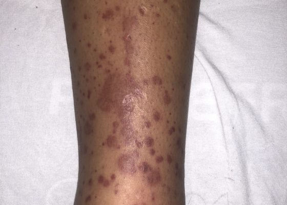
The images show a raised, palpable, purpuric rash on the lower extremities, surrounded by a mild, 1+ non-pitting edema. Several of the lesions are exfoliated with serous discharge. There is no surrounding erythema, fluctuance, or lymphangitis to suggest cellulitis. There was no tenderness to palpation; however, pruritus was exacerbated on palpation.
DermatologyCardiology/VascularVisual EM
Creative Commons images

ECG Stampede

Benjamin L Cooper, MD*, Jonathan A Giordano, DO*, Catherine E Reynolds, MD*, and Tom T Fadial, MD*

DOI: https://doi.org/10.21980/J8PP93 Issue 4:4[mrp_rating_result]
ECG Stampede is a unique educational program comprised of a comprehensive, video-based curriculum with accompanying student and instructor guides. ECG Stampede also features an engaging game accessible via modern browsers and dedicated mobile applications for iOS and Android devices. The gamified model presents learners with a batch of ECGs for which they must assign one of four triage acuities.
Cardiology/VascularInnovations
Creative Commons images

Atypical Presentation of Abdominal Aortic Aneurysm

Michael Rohinton Mirza, MD* and Christopher Bryczkowski, MD*

DOI: https://doi.org/10.21980/J82W6F Issue 4:4 [mrp_rating_result]
Bedside ultrasound revealed an abdominal aortic aneurysm (AAA) with concern for dissection vs thrombus/hematoma due to an area of echogenicity within the lumen of the vessel, since normal blood vessels (including the aorta) have lumens that are uniformly anechoic. An intimal flap concerning for dissection appears as a hyperechoic stripe within the lumen of the vessel on ultrasound, often with a hypoechoic and/or anechoic area appreciated underneath the flap, indicating a separate area of blood flow. If this visualized area is of significant size, color doppler can be used to confirm blood flow on both sides of the flap. Given his bedside ultrasound findings, the patient underwent emergent computed tomography scan and was found to have an enlarged infrarenal abdominal aortic aneurysm, with acute intramural hematoma, extending into bilateral common iliac arteries.
Cardiology/VascularVisual EM
Creative Commons images

Thoracic Aortic Aneurysm Measured by Point of Care Ultrasound Suprasternal Notch View

Hamid Ehsani-Nia, DO* and Christopher Bryczkowski, MD*

DOI: https://doi.org/10.21980/J8Z64V Issue 4:4[mrp_rating_result]
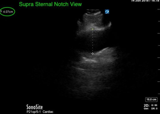
Point-of-care cardiac echocardiogram demonstrated a dilated ascending aorta (illustrated in red) measuring approximately 4 cm in the parasternal long axis (PLAX). A dilated aortic arch (illustrated in green) also measuring approximately 4 cm was appreciated using the suprasternal notch view (SSNV). A follow-up computed tomography angiogram (CTA) was performed, validating bedside ultrasound measurements.
UncategorizedCardiology/VascularVisual EM

A Story About Mesenteric Ischemia

Annahieta Kalantari, DO*

DOI: https://doi.org/10.21980/J8J33Q Issue 4:3[mrp_rating_result]
We aim to teach the presentation and management of cardiovascular emergencies through the creation of a flipped classroom design. This unique, innovative curriculum utilizes resources chosen by education faculty and resident learners, study questions, real-life experiences, and small group discussions in place of traditional lectures. In doing so, a goal of the curriculum is to encourage self-directed learning, improve understanding and knowledge retention, and improve the educational experience of our residents.
Abdominal/GastroenterologyCardiology/VascularSmall Group Learning
«‹8›»
Page 8 of 14

JETem is an online, open access, peer-reviewed, journal-repository for EM educators. We are PMC Indexed.

Most Viewed

  • Simple Auricular Hematoma (“Cauliflower Ear”) Task Trainer
  • The Three Breath Method: Teaching Coping Mechanisms for Processing Patient Death
  • Clinical Decision Making: Acute Ischemic Stroke with Large Vessel Occlusion
  • PRIMER: Premedical Introduction to Mentored Emergency Research: Connecting Undergraduate Assistants and Emergency Medicine Residents in a Community Hospital Setting
  • Dangerous Dysrhythmias in the ED: An Interactive EKG Workshop for Emergency Medicine Clerkship Learners

Visit Our Collaborators


Creative Commons Licence
This work is licensed under a Creative Commons Attribution 4.0 International License.

About

Education

Learners should benefit from active learning. JETem accepts submissions of team-based learning, small group learning, simulation, podcasts, lectures, innovations, curricula, question sets, and visualEM.

Scholarship

We believe educators should advance through the scholarship of their educational work. JETem gives educators the opportunity to publish scholarly academic work so that it may be widely distributed, thereby increasing the significance of their results.

Links

  • Home
  • Aim and Scope
  • Current Issue
  • For Reviewers
  • Instructions for Authors
  • Contact Us

Newsletter

Sign up to receive updates from JETem regarding newly published issues and findings.

Copyright Creative Commons Attribution 4.0 International