
JETem
  • Home
  • About
    • Aim and Scope
    • Our Team
    • Editorial Board
    • FAQ
  • Issues
    • Current Issue
    • Ahead of Print
    • Past Issues
  • Visual EM
    • Latest Visual EM
    • Search Visual EM
    • Thumbnail Library
  • For Authors
    • Instructions for Authors
    • Submit to JETem
    • Photo Consent
    • Policies
      • Peer Review Policy
      • Copyright Policy
      • Editorial Policy, Ethics and Responsibilities
      • Conflicts of Interest & Informed Consent
      • Open Access Policy
      • Acceptance Fee
  • For Reviewers
    • Instructions for JETem Reviewers
    • Interested in Being a JETem Reviewer?
  • Topic
    • Abdominal/Gastroenterology
    • Administration
    • Board Review
    • Cardiology/Vascular
    • Certifying Exam Cases
      • Clinical Decision-Making
      • Prioritization
      • Communication
      • Procedure & Ultrasound
    • Clinical Informatics, Telehealth and Technology
    • Dermatology
    • EMS
    • Endocrine
    • ENT
    • Faculty Development
    • Genitourinary
    • Geriatrics
    • Hematology/Oncology
    • Infectious Disease
    • Miscellaneous (stats, etc)
    • Neurology
    • Ob/Gyn
    • Ophthalmology
    • Orthopedics
    • Pediatrics
    • Pharmacology
    • Procedures
    • Psychiatry
    • Renal/Electrolytes/Nutrition
    • Respiratory
    • Social Determinants of Health
    • Toxicology
    • Trauma
    • Ultrasound
    • Urology
    • Wellness
    • Wilderness
  • Modality
    • Certifying Exam Practice Cases
      • Clinical Care Cases
      • Communication & Procedural Cases
      • Old School Oral Board Cases
    • Curricula
    • Innovations
    • Lectures
    • Podcasts
    • Simulation
    • Small Group Learning
    • Team Based Learning (TBL)
    • Visual EM
  • Contact Us

Cardiology/Vascular

Hyperkalemia ECG JETem 2016

Hyperkalemia on ECG

Bryson Hicks, MD*

DOI: https://doi.org/10.21980/J8K017Issue 1:2[mrp_rating_result]
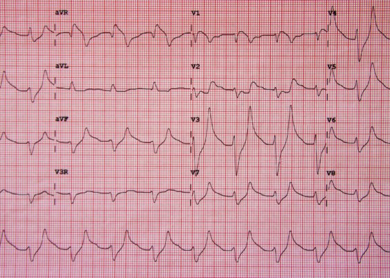
Initial ECG shows tall, peaked T waves, most prominently in V3 and V4, as well as QRS widening. These findings are consistent with hyperkalemia, which was promptly treated. Follow-up ECG post-treatment shows narrowing of the QRS complexes and normalization of peaked T waves.
Cardiology/VascularVisual EM
Traumatic Aortic Injury. Sag CT 1, Annotated. JETem 2016

Traumatic Aortic Injury

Brianna Miner*, Megan Boysen Osborn, MD, MHPE* and Shannon Toohey, MD, MA*

DOI: https://doi.org/10.21980/J85P4JIssue 1:2[mrp_rating_result]
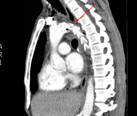
The initial chest x-ray showed an abnormal superior mediastinal contour (blue line), suggestive of a possible aortic injury. The CT angiogram showed extensive circumferential irregularity and outpouching of the distal aortic arch (red arrows) compatible with aortic transection. In addition, there was a circumferential intramural hematoma, which extended through the descending aorta to the proximal infrarenal abdominal aorta (green arrow). There was also an extensive surrounding mediastinal hematoma extending around the descending aorta and supraaortic branches (purple arrows).
TraumaCardiology/VascularVisual EM
WPW EKG 1, initial EKG. JETem 2016

Wolff-Parkinson-White Syndrome: Electrocardiogram

Brianna Miner*, Jonathan Patane, MD*, and Carrie Chandwani, MD*

DOI: https://doi.org/10.21980/J8201KIssue 1:2[mrp_rating_result]
The initial EKG showed wide complex, irregular tachycardia > 200 bpm (EKG 1). Given the possibility of Wolff-Parkinson-White (WPW), procainamide was given to the patient. The patient’s heart rate responded and decreased to 120-140 bpm with narrowing of the QRS complex. A repeat EKG showed narrow complex tachycardia without P waves approximately 120 bpm (EKG 2). Once the procainamide infusion was complete, the patient had converted to sinus rhythm with a delta wave now apparent, consistent with WPW (EKG 3).
Cardiology/VascularVisual EM
Creative Commons images

Atrial Myxoma

Alisa Wray, MD*

DOI: https://doi.org/10.21980/J87P45Issue 1:1[mrp_rating_result]
Bedside ultrasound revealed the presence of a left atrial mass that appeared to be tethered to the mitral valve. The mass was best viewed on ultrasound in the apical four-chamber window with the phased array probe placed over the patients’ point of maximal impact (PMI), with the patient in left lateral decubitus position.
UltrasoundCardiology/VascularVisual EM
Creative Commons images

Acute Pericarditis: Electrocardiogram

Jason Mefford, MD* and Shannon L Toohey, MD*

DOI: https://doi.org/10.21980/J8059QIssue 1:1[mrp_rating_result]
The ECG shows diffuse ST- elevation. The patient also has mild PR-depression, most notably in the inferior and lateral leads. The patient also has minimal PR elevation in lead aVR. The patient was diagnosed with acute pericarditis (ECG stage 1).
Cardiology/VascularVisual EM
Creative Commons images

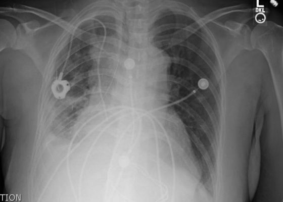
Pericardial Effusion CXR

[mrp_rating_result]
  Keywords: radiology, x-ray, oncology, pericardial effusion, cardiology, CXR
Cardiology/Vascular
Creative Commons images

Raynaud’s Phenomenon

[mrp_rating_result]
  Photograph contributed by Richard Saunders‎, DO, FACEP Keywords: Raynaud’s phenomenon, Raynaud’s syndrome.
Cardiology/VascularDermatology
Creative Commons images

Congestive Heart Failure X-Ray

[mrp_rating_result]
  Keywords: radiology, x-ray, congestive heart failure, CHF, cardiology, CXR
Cardiology/Vascular
«‹14
Page 14 of 14

JETem is an online, open access, peer-reviewed, journal-repository for EM educators. We are PMC Indexed.

Most Viewed

  • Simple Auricular Hematoma (“Cauliflower Ear”) Task Trainer
  • The Three Breath Method: Teaching Coping Mechanisms for Processing Patient Death
  • Clinical Decision Making: Acute Ischemic Stroke with Large Vessel Occlusion
  • PRIMER: Premedical Introduction to Mentored Emergency Research: Connecting Undergraduate Assistants and Emergency Medicine Residents in a Community Hospital Setting
  • Dangerous Dysrhythmias in the ED: An Interactive EKG Workshop for Emergency Medicine Clerkship Learners

Visit Our Collaborators


Creative Commons Licence
This work is licensed under a Creative Commons Attribution 4.0 International License.

About

Education

Learners should benefit from active learning. JETem accepts submissions of team-based learning, small group learning, simulation, podcasts, lectures, innovations, curricula, question sets, and visualEM.

Scholarship

We believe educators should advance through the scholarship of their educational work. JETem gives educators the opportunity to publish scholarly academic work so that it may be widely distributed, thereby increasing the significance of their results.

Links

  • Home
  • Aim and Scope
  • Current Issue
  • For Reviewers
  • Instructions for Authors
  • Contact Us

Newsletter

Sign up to receive updates from JETem regarding newly published issues and findings.

Copyright Creative Commons Attribution 4.0 International