
Cardiology/Vascular

A Case Report of a Transected Carotid Artery Caused by a Stab Wound to the Neck
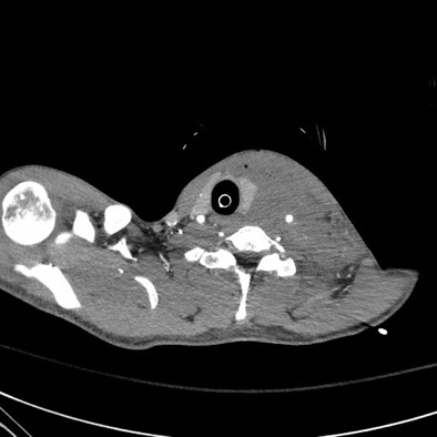
DOI: https://doi.org/10.21980/J8BP8MThe post intubation chest x-ray (CXR) showed severe rightward displacement of the trachea (purple arrow). The computed tomography angiogram (CTA) showed transection of the left common carotid artery (LCCA), extensive neck hematoma without extravasation and severe tracheal deviation to the right (blue arrow). The intravenous (IV) contrasted chest computed tomography (CT) image showed a lateral contrast projection from the aortic arch at the level of the isthmus (green and pink arrows). There were no other significant injuries reported on the CT scans of the chest, abdomen and pelvis.

Extracorporeal Membrane Oxygenation (ECMO) for Refractory Cardiac Arrest
DOI: https://doi.org/10.21980/J88W69ABSTRACT: Audience: Our target audience includes emergency medicine residents/physicians. Introduction: Treating cardiac arrest is a common theme during simulated emergency medicine training; however, less time is focused on treating refractory cases of cardiac arrest. There are varying definitions of refractory cardiac arrest, but it is most commonly defined as the inability to obtain return of spontaneous circulation (ROSC) after 10-30
Cardiac Tamponade
DOI: https://doi.org/10.21980/J81D1DBy the end of this simulation session, the learner will be able to: (1) describe a diagnostic differential for dizziness (2) describe the pathophysiology of cardiac tamponade (3) describe the acute management of cardiac tamponade, including fluid bolus and pericardiocentesis (4) describe the electrocardiogram (ECG) findings of pericardial effusion (5) describe the ultrasound findings of cardiac tamponade (6) describe the indications for emergent bedside pericardiocentesis versus medical stabilization and delayed pericardiocentesis for cardiac tamponade (7) describe the procedural steps for pericardiocentesis, and (8) describe your state’s laws regarding disclosure for sentinel events.
Paroxysmal Ventricular Standstill—A Case Report of all Ps and no QRS in Ventricular Asystole
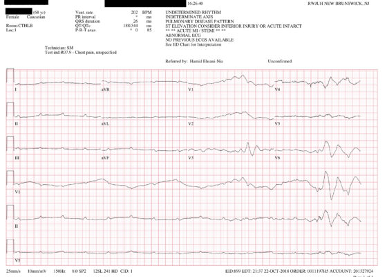
DOI: https://doi.org/10.21980/J8SS79In route, it was proposed that this patient was suffering from a dysrhythmia due to the transient episodes of syncope with lack of ventricular activity on telemetry. Upon close examination of the rhythm strips as well as the ECG, P waves can be visualized without any accompanying QRS complexes lasting multiple seconds (ED ECG blue arrows). Additionally, the rhythm has an intrinsic rate of 100 beats per minute and has a consistent morphology with no evidence of ventricular activity due to the lack of QRS complexes. As a result, the rhythm likely originates in the atria with no passage of impulses into the ventricles through the atrioventricular (AV) node versus an accelerated ventricular rhythm where QRS complexes would be seen.8 These rhythm strips demonstrate an example of VS. There is preserved native atrial automaticity, with an intact sinoatrial (SA) node, with a complete lack of ventricular electrical activity

Severe Hyperkalemia
DOI: https://doi.org/10.21980/J8KH1DThe initial ECG obtained upon arrival shows what is commonly referred to as a sine wave pattern. This patient does have a biventricular pacemaker which would ordinarily create a wide QRS complex mimicking an intraventricular conduction delay. However, the QRS complex here is exceptionally wide, in excess of 400 milliseconds (normal: less than 120 milliseconds). As the QRS widens, alongside other deflections present on the ECG, it morphologically mimics a mathematical sine wave.

Caught on CT! The Case of the Hemodynamically Stable Ruptured Abdominal Aortic Aneurysm
DOI: https://doi.org/10.21980/J8B07BThe associated images demonstrate the transverse, sagittal, and coronal views of a 6.8 cm infrarenal ruptured AAA continuous with a 4 cm right common iliac aneurysm (transverse, sagittal and coronal). Active hemorrhage was seen contained within the aortic wall, and retroperitoneal bleeding can be appreciated with asymmetric enlargement of the left psoas muscle (coronal - red arrow).1 Plaque and calcifications with a residual opacified true lumen is also present (transverse – red star, sagittal – red arrow). Known as the tangential calcium sign, this is a common radiologic finding of AAAs.2
Post-Coital Sudden Cardiac Arrest Due to Non-Traumatic Subarachnoid Hemorrhage—A Case Report
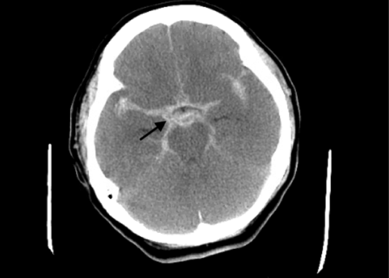
DOI: https://doi.org/10.21980/J8663NThe electrocardiogram demonstrated sinus tachycardia with ST segment elevation in lead aVR (black arrows) and diffuse ST depressions concerning for possible ST elevation myocardial infarction (STEMI). Given the events reported and the patient’s neurologic exam without sedation, non-contrast CT of the head was ordered; imaging showed evidence of a large subarachnoid hemorrhage, mostly at the level of the Circle of Willis (black arrow) concerning for an aneurysmal bleed as well as mild generalized white matter density suggestive of cerebral edema.

Ascending Thoracic Aortic Dissection: A Case Report of Rapid Detection Via Emergency Echocardiography with Suprasternal Notch Views
DOI: https://doi.org/10.21980/J8WW6WVideo of parasternal long-axis bedside transthoracic echocardiogram: The initial images showed grossly normal left ventricular function, and no pericardial effusion or evidence of cardiac tamponade. However, the proximal aorta beyond the aortic valve was poorly-visualized in this window.