
JETem
  • Home
  • About
    • Aim and Scope
    • Our Team
    • Editorial Board
    • FAQ
  • Issues
    • Current Issue
    • Ahead of Print
    • Past Issues
  • Visual EM
    • Latest Visual EM
    • Search Visual EM
    • Thumbnail Library
  • For Authors
    • Instructions for Authors
    • Submit to JETem
    • Photo Consent
    • Policies
      • Peer Review Policy
      • Copyright Policy
      • Editorial Policy, Ethics and Responsibilities
      • Conflicts of Interest & Informed Consent
      • Open Access Policy
      • Acceptance Fee
  • For Reviewers
    • Instructions for JETem Reviewers
    • Interested in Being a JETem Reviewer?
  • Topic
    • Abdominal/Gastroenterology
    • Administration
    • Board Review
    • Cardiology/Vascular
    • Certifying Exam Cases
      • Clinical Decision-Making
      • Prioritization
      • Communication
      • Procedure & Ultrasound
    • Clinical Informatics, Telehealth and Technology
    • Dermatology
    • EMS
    • Endocrine
    • ENT
    • Faculty Development
    • Genitourinary
    • Geriatrics
    • Hematology/Oncology
    • Infectious Disease
    • Miscellaneous (stats, etc)
    • Neurology
    • Ob/Gyn
    • Ophthalmology
    • Orthopedics
    • Pediatrics
    • Pharmacology
    • Procedures
    • Psychiatry
    • Renal/Electrolytes/Nutrition
    • Respiratory
    • Social Determinants of Health
    • Toxicology
    • Trauma
    • Ultrasound
    • Urology
    • Wellness
    • Wilderness
  • Modality
    • Certifying Exam Practice Cases
      • Clinical Care Cases
      • Communication & Procedural Cases
      • Old School Oral Board Cases
    • Curricula
    • Innovations
    • Lectures
    • Podcasts
    • Simulation
    • Small Group Learning
    • Team Based Learning (TBL)
    • Visual EM
  • Contact Us

Cardiology/Vascular

Creative Commons images

Osborn Waves

James Elliot Karz, DO*, Grant Wei, MD* and Chirag Shah, MD*

DOI: https://doi.org/10.21980/J8G34G Issue 3:2[mrp_rating_result]
The initial ECG shows a junctional rhythm with Osborn waves (or J point elevations/J waves) in the lateral precordial leads, as well as the limb leads (Image 1). The second ECG, 49 minutes later, shows an improving ventricular rate and Osborn wave height decrease of approximately 50% (Image 2). 
Cardiology/VascularVisual EM
Creative Commons images

Elderly female with acute abdominal pain presenting with Superior Mesenteric Artery Thrombus

Sassan Ghassemzadeh, MD*

DOI: https://doi.org/10.21980/J82W52 Issue 3:2[mrp_rating_result]
Computed tomography (CT) angiogram of the abdomen and pelvis revealed a superior mesenteric artery (SMA) thrombosis 5 cm from the origin off of the abdominal aorta. As seen in the sagittal view, there does not appear to be any contrast 5 cm past the origin of the SMA. On the axial views, you can trace the SMA until the point that there is no longer any contrast visible which indicates the start of the thrombus.  The SMA does not appear to be reconstituted. There was normal flow to the celiac artery. (See annotated images).
Abdominal/GastroenterologyCardiology/VascularVisual EM
Creative Commons images

Tricuspid Annular Plane Systolic Excursion (TAPSE) in a Patient with Pulmonary Emboli

Nicole Zawada, BS*, Ethan Kunstadt, MD* and Maili Alvarado, MD^

DOI: https://doi.org/10.21980/J8M052 Issue 3:1[mrp_rating_result]
Video 1 and Image 1 show a thrombus in the patient’s IVC. Video 2 and Images 2-3 demonstrate a positive TAPSE of less than 17mm (blue arrow length) with a significantly dilated RV, indicating abnormal excursion of the tricuspid annulus and right ventricular dysfunction.  
RespiratoryCardiology/VascularVisual EM
Creative Commons images

Management of Wolff-Parkinson-White Tachyarrhythmia Presenting as Syncope with Seizure-like Activity

Samuel Kaplan, BS* and Lindsey Spiegelman, MD*

DOI: https://doi.org/10.21980/J8534P Issue 2:4[mrp_rating_result]
At the end of this simulation session the learner will: 1) Recognize clinical history suggestive of cardiogenic syncope, 2) recognize clinical evidence of Wolff-Parkinson-White syndrome, 3) promptly and appropriately treat unstable WPW tachyarrhythmia.
Cardiology/VascularSimulation
Creative Commons images

Asymptomatic Wolff-Parkinson-White Syndrome: Incidental EKG

Samer Assaf, MD* and Christopher Libby, MD, MPH^

DOI: https://doi.org/10.21980/J8T05X Issue 2:3[mrp_rating_result]
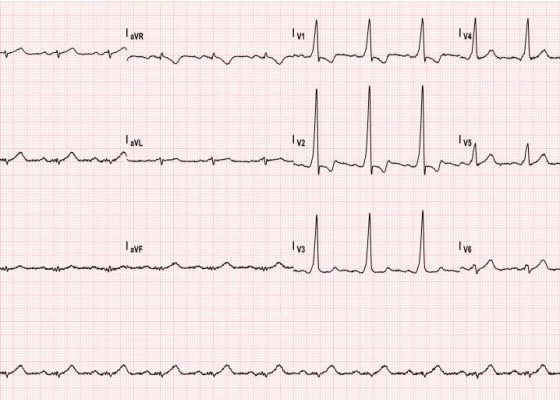
The ECG shows slurred up-stroking of the QRS complexes characteristic of a delta wave. The PR interval is normal; however, the QT interval is greater than 110ms.
Cardiology/VascularVisual EM
Creative Commons images

Renal Infarction from Type B Aortic Dissection

Marit Tweet, MD* and James Roy Waymack, MD*

DOI: https://doi.org/10.21980/J8HG9G Issue 2:3[mrp_rating_result]
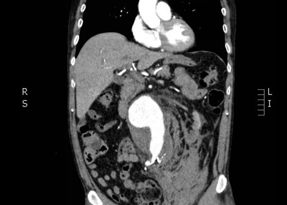
Initial abdominal images demonstrated a dissection flap; therefore, a CTA of the chest was also obtained. These images revealed a Stanford type B aortic dissection beginning just distal to the left subclavian artery and extending to the origin of the inferior mesenteric artery. The right renal artery arose from the true lumen of the dissection while the left renal artery arose from the false lumen. This case is interesting as imaging shows the lack of perfusion to the left kidney, residing in the retroperitoneum, which correlates with her non-descript abdominal and left flank pain.
Cardiology/VascularRenal/ElectrolytesVisual EM
Creative Commons images

Ruptured Abdominal Aortic Aneurysm

Jessica Andrusaitis, BS, MS* and Jonathan Peña, MD*

DOI: https://doi.org/10.21980/J8FP6SIssue 2:3[mrp_rating_result]
CTA demonstrated a ruptured 7.4 cm infrarenal abdominal aortic aneurysm with a large left retroperitoneal hematoma.
Cardiology/VascularVisual EM
Creative Commons images

Use of Bedside Compression Ultrasonography for Diagnosis of Deep Venous Thrombosis

Mohamad Moussa, MD* and Maher Abdo, BS*

DOI: https://doi.org/10.21980/J81G94 Issue 2:3[mrp_rating_result]
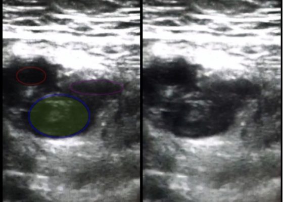
As shown in the still image of the performed ultrasound, a transverse view of the proximal-thigh revealed a visible thrombus (green shading) occluding the lumen of the left common femoral vein (blue ring), which was non-compressible when direct pressure was applied to the probe. Also visible is a patent and compressible branch of the common femoral vein (purple ring) and the femoral artery (red ring), highlighted by its thick vessel wall and pulsatile motion.
UltrasoundCardiology/VascularVisual EM
«‹12›»
Page 12 of 14

JETem is an online, open access, peer-reviewed, journal-repository for EM educators. We are PMC Indexed.

Most Viewed

  • Simple Auricular Hematoma (“Cauliflower Ear”) Task Trainer
  • The Three Breath Method: Teaching Coping Mechanisms for Processing Patient Death
  • Clinical Decision Making: Acute Ischemic Stroke with Large Vessel Occlusion
  • PRIMER: Premedical Introduction to Mentored Emergency Research: Connecting Undergraduate Assistants and Emergency Medicine Residents in a Community Hospital Setting
  • Dangerous Dysrhythmias in the ED: An Interactive EKG Workshop for Emergency Medicine Clerkship Learners

Visit Our Collaborators


Creative Commons Licence
This work is licensed under a Creative Commons Attribution 4.0 International License.

About

Education

Learners should benefit from active learning. JETem accepts submissions of team-based learning, small group learning, simulation, podcasts, lectures, innovations, curricula, question sets, and visualEM.

Scholarship

We believe educators should advance through the scholarship of their educational work. JETem gives educators the opportunity to publish scholarly academic work so that it may be widely distributed, thereby increasing the significance of their results.

Links

  • Home
  • Aim and Scope
  • Current Issue
  • For Reviewers
  • Instructions for Authors
  • Contact Us

Newsletter

Sign up to receive updates from JETem regarding newly published issues and findings.

Copyright Creative Commons Attribution 4.0 International