
JETem
  • Home
  • About
    • Aim and Scope
    • Our Team
    • Editorial Board
    • FAQ
  • Issues
    • Current Issue
    • Ahead of Print
    • Past Issues
  • Visual EM
    • Latest Visual EM
    • Search Visual EM
    • Thumbnail Library
  • For Authors
    • Instructions for Authors
    • Submit to JETem
    • Photo Consent
    • Policies
      • Peer Review Policy
      • Copyright Policy
      • Editorial Policy, Ethics and Responsibilities
      • Conflicts of Interest & Informed Consent
      • Open Access Policy
      • Acceptance Fee
  • For Reviewers
    • Instructions for JETem Reviewers
    • Interested in Being a JETem Reviewer?
  • Topic
    • Abdominal/Gastroenterology
    • Administration
    • Board Review
    • Cardiology/Vascular
    • Certifying Exam Cases
      • Clinical Decision-Making
      • Prioritization
      • Communication
      • Procedure & Ultrasound
    • Clinical Informatics, Telehealth and Technology
    • Dermatology
    • EMS
    • Endocrine
    • ENT
    • Faculty Development
    • Genitourinary
    • Geriatrics
    • Hematology/Oncology
    • Infectious Disease
    • Miscellaneous (stats, etc)
    • Neurology
    • Ob/Gyn
    • Ophthalmology
    • Orthopedics
    • Pediatrics
    • Pharmacology
    • Procedures
    • Psychiatry
    • Renal/Electrolytes/Nutrition
    • Respiratory
    • Social Determinants of Health
    • Toxicology
    • Trauma
    • Ultrasound
    • Urology
    • Wellness
    • Wilderness
  • Modality
    • Certifying Exam Practice Cases
      • Clinical Care Cases
      • Communication & Procedural Cases
      • Old School Oral Board Cases
    • Curricula
    • Innovations
    • Lectures
    • Podcasts
    • Simulation
    • Small Group Learning
    • Team Based Learning (TBL)
    • Visual EM
  • Contact Us

Cardiology/Vascular

Creative Commons images

Don’t Forget the Pacemaker – A Rare Complication

Amanda Esposito, MD*, Kenneth Rapp, MD* and Albert Ritter, MD*

DOI: https://doi.org/10.21980/J8GS7HIssue 3:4[mrp_rating_result]
The ECG demonstrated the presence of pacemaker spikes without appropriate capture (green arrows) and a ventricular escape rhythm which can be identified by an absence of P waves prior to the QRS complex (purple arrows). The portable chest X- demonstrated displaced pacemaker leads (red arrows) that were coiled around the pulse generator (blue arrow).   
Cardiology/VascularVisual EM

Novel Emergency Medicine Curriculum Utilizing Self-Directed Learning and the Flipped Classroom Method: Cardiovascular Emergencies Small Group Module

Michael Barrie, MD*, Erin Wenzel, MD*, Colin Kaide, MD*, Daniel Bachmann, MD*, Daniel Martin, MD, MBA*, Jennifer Mitzman MD*^, Benjamin Ostro, MD*, Beth Bubolz, MD^, Kristin Stukus, MD^, Farhad Aziz, MD*, Cynthia Leung, MD*, Howard Werman, MD*, Alyssa Tyransky* and Andrew King, MD*

DOI: https://doi.org/10.21980/J8X334 Issue 3:3[mrp_rating_result]
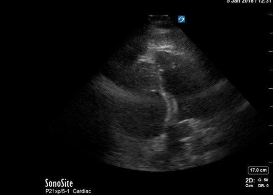
We aim to teach the presentation and management of cardiovascular emergencies through the creation of a flipped classroom design. This unique, innovative curriculum utilizes resources chosen by education faculty and resident learners, study questions, real-life experiences, and small group discussions in place of traditional lectures. In doing so, a goal of the curriculum is to encourage self-directed learning, improve understanding and knowledge retention, and improve the educational experience of our residents.
CurriculumCardiology/Vascular
Creative Commons images

Right Ventricular Dilation in Patient With Submassive Pulmonary Embolism

Adrian Diez, MD* and Christopher Bryczkowski, MD*

DOI: https://doi.org/10.21980/J82P84Issue 3:3[mrp_rating_result]
Bedside echocardiography four chamber view revealed enlarged right ventricular (RV) to left ventricular (LV) ratio (greater than 1) on apical four-chamber view (see red and blue outlines respectively). The right atrium is not clearly delineated in this image and therefore is not outlined. One can also rule out a large pericardial effusion as the cause of her dyspnea, since there is no large hypoechoic collection surrounding the heart on either four- chamber view or parasternal long view.
Cardiology/VascularVisual EM

Fainting Spells

Brittany Guest, DO*, Amir Rouhani, MD* and Steven Lai, MD*

DOI: https://doi.org/10.21980/J8Z91R Issue 3:2[mrp_rating_result]
ABSTRACT: Audience: The target audience for this simulation is 4th year medical students, emergency medicine residents, pediatric residents, and family medicine residents. Introduction: Brugada syndrome is defined as the combination of specific electrocardiogram (ECG) changes and clinical manifestations of a ventricular arrhythmia, including syncope and sudden cardiac arrest.1 Brugada syndrome is caused by a mutation in the phase-0 cardiac sodium channel. This
Cardiology/VascularSimulation

Chest Pain with Acute Coronary Syndrome

Alisa Wray, MD*

DOI: https://doi.org/10.21980/J8K04C Issue 3:2[mrp_rating_result]
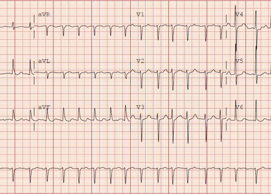
At the end of this case learners should be able to prepare a differential for patients with chest pain, provide appropriate medications for a patient with chest pain, recognize an ST segment elevation MI, and appropriately disposition a patient with acute myocardial infarction (AMI).
Cardiology/VascularSimulation
Creative Commons images

Osborn Waves in a Severely Hypothermic Patient

Jon Van Heukelom, MD*

DOI: https://doi.org/10.21980/J8H34SIssue 3:2[mrp_rating_result]
The initial EKG shows marked elevation of the J-point (point where the QRS segment joins the ST segment), otherwise known as an “Osborn Wave” (see black arrows).  A subsequent EKG obtained after active rewarming, showed resolution of the Osborn waves.
Cardiology/VascularVisual EM
Creative Commons images

Torsades de Pointes

Richard J Chen, MD*, Grant Wei, MD* and Chirag Shah, MD*

DOI: https://doi.org/10.21980/J87K91 Issue 3:2[mrp_rating_result]
The patient was found to be in a polymorphic ventricular tachycardia; he was alert, awake and asymptomatic. A rhythm strip showed a wide complex tachycardia with the QRS complex varying in amplitude around the isoelectric line consistent with Torsades de Pointes.
Cardiology/VascularVisual EM
Creative Commons images

Type 1 Brugada Syndrome

Sha Yan, DO*, Grant Wei, MD* and Chirag Shah, MD*

DOI: https://doi.org/10.21980/J8V91T Issue 3:2[mrp_rating_result]
ECG shows an incomplete right bundle branch block (blue arrow) with coved ST segment elevation and an inverted T wave in V1 (red arrow) and ST segment elevation in V2 (black arrow).
Cardiology/VascularVisual EM
«‹11›»
Page 11 of 14

JETem is an online, open access, peer-reviewed, journal-repository for EM educators. We are PMC Indexed.

Most Viewed

  • Simple Auricular Hematoma (“Cauliflower Ear”) Task Trainer
  • The Three Breath Method: Teaching Coping Mechanisms for Processing Patient Death
  • Clinical Decision Making: Acute Ischemic Stroke with Large Vessel Occlusion
  • PRIMER: Premedical Introduction to Mentored Emergency Research: Connecting Undergraduate Assistants and Emergency Medicine Residents in a Community Hospital Setting
  • Dangerous Dysrhythmias in the ED: An Interactive EKG Workshop for Emergency Medicine Clerkship Learners

Visit Our Collaborators


Creative Commons Licence
This work is licensed under a Creative Commons Attribution 4.0 International License.

About

Education

Learners should benefit from active learning. JETem accepts submissions of team-based learning, small group learning, simulation, podcasts, lectures, innovations, curricula, question sets, and visualEM.

Scholarship

We believe educators should advance through the scholarship of their educational work. JETem gives educators the opportunity to publish scholarly academic work so that it may be widely distributed, thereby increasing the significance of their results.

Links

  • Home
  • Aim and Scope
  • Current Issue
  • For Reviewers
  • Instructions for Authors
  • Contact Us

Newsletter

Sign up to receive updates from JETem regarding newly published issues and findings.

Copyright Creative Commons Attribution 4.0 International