X-Ray
Traumatic Aortic Injury
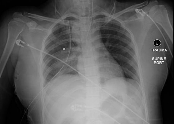
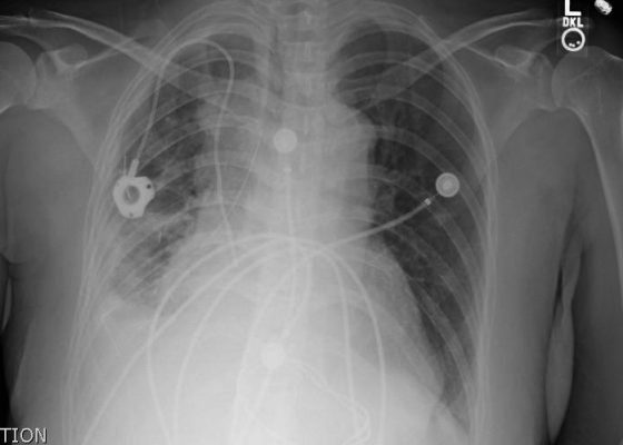
DOI: https://doi.org/10.21980/J85P4JThe initial chest x-ray showed an abnormal superior mediastinal contour (blue line), suggestive of a possible aortic injury. The CT angiogram showed extensive circumferential irregularity and outpouching of the distal aortic arch (red arrows) compatible with aortic transection. In addition, there was a circumferential intramural hematoma, which extended through the descending aorta to the proximal infrarenal abdominal aorta (green arrow). There was also an extensive surrounding mediastinal hematoma extending around the descending aorta and supraaortic branches (purple arrows).
Posterior Elbow Dislocation
DOI: https://doi.org/10.21980/J8X593Elbow dislocations are classified by the position of the radio-ulnar joint relative to the humerus.1 Images 1, 2, and 3 show a left posterior elbow dislocation; the radius and ulna (red lines) are displaced posteriorly with respect to the distal humerus (blue line). The lateral view of the elbow most clearly shows this: trochlear notch of the ulna (red line) is empty and displaced posteriorly relative to the trochlea (blue line). There is no associated fracture. Images 4 and 5 show the elbow status-post reduction, demonstrating proper alignment of the distal humerus (blue line) with the radius and ulna (red lines).
Open Dislocation of Fifth Digit
DOI: https://doi.org/10.21980/J8J01XPhysical exam revealed an open dislocation of the proximal interphalangeal joint (PIP) of the right fifth digit. X-ray confirmed dislocation and revealed no fractures. The patient received a tetanus booster, Cefazolin, and the dislocation was then washed out and reduced. Multiple reduction attempts were made and were only successful once the metacarpophalangeal joints were held in 90 degree flexion, which relaxed the lateral bands and enabled the finger to be reduced.
Large Right Pleural Effusion
DOI: https://doi.org/10.21980/J8D59FChest x-ray and bedside ultrasound revealed a large right pleural effusion, estimated to be greater than two and a half liters in size.
Hampton’s Hump in Pulmonary Embolism
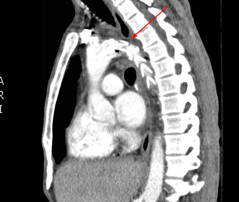
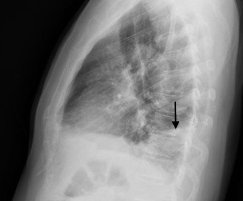
DOI: https://doi.org/10.21980/J83W27In the lateral view chest x-ray, there is a Hampton’s Hump, a pleural based, wedge-shaped opacity at the base of the right lung, representing lung infarction (black arrow). These findings correlate with the sagittal view on CT angiography of the chest. The CT chest also shows a filling defect in the distal posterior basal segmental pulmonary artery (white arrow), as demonstrated by the absence of contrast enhancement in the distal portion of the vessel. This is associated with an opacification of the lung parenchyma distal to the occlusion (red arrow), representing lung infarction.
Normal CXR: AP and Lateral
Keywords: radiology, normal, chest, CXR, pulm, respiratory, cardiovascular, CV, AP, lateral
Normal CXR and Post-Intubation CXR
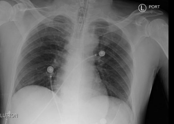
Keywords: radiology, normal, intubation, CXR, chest, respiratory, respiratory failure, AP, ETT, post-intubation
Hemothorax Pre and Post Chest Tube CXR
Keywords: radiology, x-ray, trauma, hemothorax, chest tube, tube thoracostomy, respiratory, pulmonary
Pericardial Effusion CXR
Keywords: radiology, x-ray, oncology, pericardial effusion, cardiology, CXR

