
JETem
  • Home
  • About
    • Aim and Scope
    • Our Team
    • Editorial Board
    • FAQ
  • Issues
    • Current Issue
    • Ahead of Print
    • Past Issues
  • Visual EM
    • Latest Visual EM
    • Search Visual EM
    • Thumbnail Library
  • For Authors
    • Instructions for Authors
    • Submit to JETem
    • Photo Consent
    • Policies
      • Peer Review Policy
      • Copyright Policy
      • Editorial Policy, Ethics and Responsibilities
      • Conflicts of Interest & Informed Consent
      • Open Access Policy
      • Acceptance Fee
  • For Reviewers
    • Instructions for JETem Reviewers
    • Interested in Being a JETem Reviewer?
  • Topic
    • Abdominal/Gastroenterology
    • Administration
    • Board Review
    • Cardiology/Vascular
    • Certifying Exam Cases
      • Clinical Decision-Making
      • Prioritization
      • Communication
      • Procedure & Ultrasound
    • Clinical Informatics, Telehealth and Technology
    • Dermatology
    • EMS
    • Endocrine
    • ENT
    • Faculty Development
    • Genitourinary
    • Geriatrics
    • Hematology/Oncology
    • Infectious Disease
    • Miscellaneous (stats, etc)
    • Neurology
    • Ob/Gyn
    • Ophthalmology
    • Orthopedics
    • Pediatrics
    • Pharmacology
    • Procedures
    • Psychiatry
    • Renal/Electrolytes/Nutrition
    • Respiratory
    • Social Determinants of Health
    • Toxicology
    • Trauma
    • Ultrasound
    • Urology
    • Wellness
    • Wilderness
  • Modality
    • Certifying Exam Practice Cases
      • Clinical Care Cases
      • Communication & Procedural Cases
      • Old School Oral Board Cases
    • Curricula
    • Innovations
    • Lectures
    • Podcasts
    • Simulation
    • Small Group Learning
    • Team Based Learning (TBL)
    • Visual EM
  • Contact Us

Ultrasound

Creative Commons images

Point-of-care Ultrasound Detection of Endophthalmitis

James Tucker MD, PhD*, Jonathan Patane, MD^ and Shadi Lahham, MD, MS^

DOI: https://doi.org/10.21980/J8G634 Issue 3:1[mrp_rating_result]
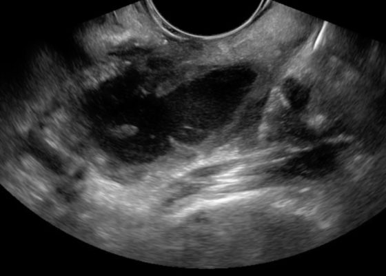
The patient’s ultrasound revealed an attached retina and a complex network of hyperechoic, mobile, membranous material in the posterior segment.
OphthalmologyUltrasoundVisual EM
Creative Commons images

Bedside Ultrasound for the Diagnosis of Peritonsillar Abscess

Harshal Bhakta, BS,* Maili Alvarado, MD^ and Daryn Towle, MD*

DOI: https://doi.org/10.21980/J8N33N Issue 2:4[mrp_rating_result]
The first video is an intraoral ultrasound using the high frequency endocavitary probe demonstrating an anechoic fluid collection adjacent to the patient’s enlarged left tonsil. The second video shows real-time ultrasound-guided successful drainage of the PTA.
UltrasoundENTInfectious DiseaseVisual EM
Creative Commons images

An Elderly Male with Amyand’s Hernia

Saema Said, MD* and Roozbeh Houshyar, MD^

DOI: https://doi.org/10.21980/J80D13 Issue 2:4[mrp_rating_result]
Ultrasound of the right scrotum shows a right inguinal hernia with an air-containing loop of bowel (white arrow) and a non-compressible appendix (yellow arrow). Coronal and axial views of abdomen-pelvis CT show a right inguinal hernia containing a loop of small bowel (white arrow) and appendix (yellow arrow).
Abdominal/GastroenterologyVisual EM
Creative Commons images

Point-of-care Ultrasound for the Diagnosis of Ectopic Pregnancy

Ahmed Farhat, BS*, Jessica Hoffmann, MD* and Maili Alvarado, MD^

DOI: https://doi.org/10.21980/J8VK7VIssue 2:4[mrp_rating_result]
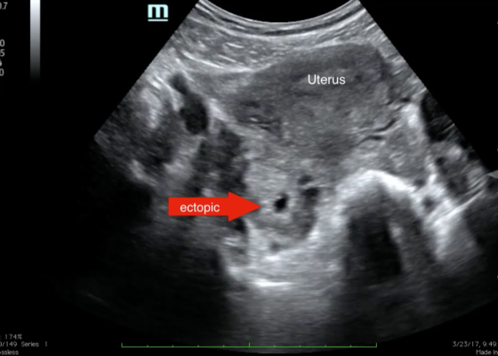
The transabdominal pelvic ultrasound shows an empty uterus (annotated) with free fluid and a right sided extrauterine gestational sac representing an ectopic pregnancy (red arrow).
Ob/GynUltrasoundVisual EM
Creative Commons images

Bedside Ultrasound for the Diagnosis of Small Bowel Obstruction

Alexander Anshus, BS* and Maili Alvarado, MD^

DOI: https://doi.org/10.21980/J86W6PIssue 2:4[mrp_rating_result]
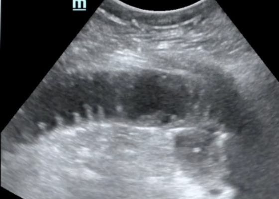
The POCUS utilizing the low frequency curvilinear probe demonstrates fluid-filled, dilated bowel loops greater than 2.5cm with to-and-fro peristalsis, and thickened bowel walls greater than 3mm, concerning for SBO. 
Abdominal/GastroenterologyVisual EM
Creative Commons images

Using Bedside Ultrasound to Rapidly Differentiate Shock

Mohamad Moussa, MD*, Jacob Willen, BS* and Sudipa Biswas, BS, MS*

DOI: https://doi.org/10.21980/J8S047 Issue 2:3[mrp_rating_result]
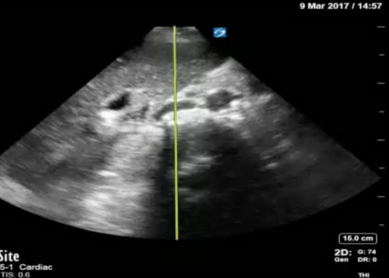
A RUSH exam demonstrated hyperdynamic cardiac contractility and collapse of the inferior vena cava (IVC) with probe compression more than 50% suggesting hypovolemia likely secondary to sepsis. Incidentally, Morrison’s pouch revealed a large right renal cyst but no signs of free fluid. A computed tomography of abdomen/pelvis showed a 10.8 x 9.5 cm right renal cyst and left lower lobe pneumonia.
UltrasoundInfectious DiseaseVisual EM
Creative Commons images

Pneumonia Diagnosed by Point-of-Care Ultrasound

Akash Patel, BA*, Steven Bunch, MD^, Maili Alvarado, MD^, Shadi Lahham, MD^ and J. Christian Fox, MD^

DOI: https://doi.org/10.21980/J8XP64 Issue 2:3[mrp_rating_result]
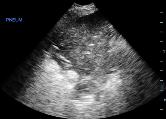
Point-of-care ultrasound of the left lower lobe demonstrates lung hepatization, a classic finding for pneumonia. In addition, a shred sign is present with both air bronchograms and focal B-lines—all suggestive of poorly aerated, consolidated lung. The patient was started on antibiotics and admitted to the hospital with a diagnosis of community-acquired pneumonia.
UltrasoundRespiratoryVisual EM
Creative Commons images

Perforated Gastric Ulcer with Intra-abdominal Abscess

Leslie Palmerlee, MD, MPH*, Scott Mackey, DO* and Michael Petrauskis, MD, MEd*

DOI: https://doi.org/10.21980/J82H0C Issue 2:3[mrp_rating_result]
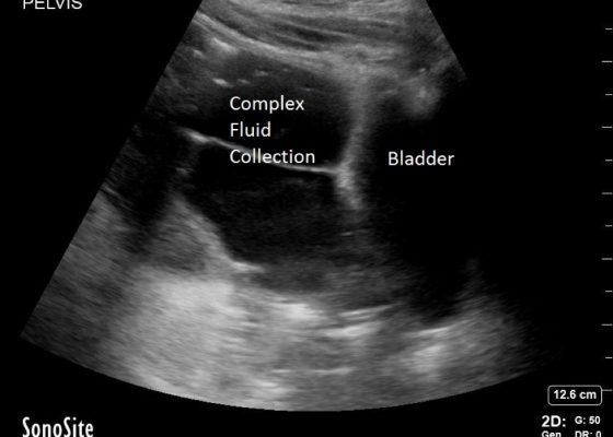
Bedside ultrasound revealed a large volume of free fluid in the right upper quadrant and in the pelvis. The fluid appeared complex with multiple septations. Its appearance was not consistent with ascites or acute intra-abdominal free fluid due to striations and pockets.
Abdominal/GastroenterologyVisual EM
«‹9›»
Page 9 of 11

JETem is an online, open access, peer-reviewed, journal-repository for EM educators. We are PMC Indexed.

Most Viewed

  • Simple Auricular Hematoma (“Cauliflower Ear”) Task Trainer
  • The Three Breath Method: Teaching Coping Mechanisms for Processing Patient Death
  • Clinical Decision Making: Acute Ischemic Stroke with Large Vessel Occlusion
  • PRIMER: Premedical Introduction to Mentored Emergency Research: Connecting Undergraduate Assistants and Emergency Medicine Residents in a Community Hospital Setting
  • Dangerous Dysrhythmias in the ED: An Interactive EKG Workshop for Emergency Medicine Clerkship Learners

Visit Our Collaborators


Creative Commons Licence
This work is licensed under a Creative Commons Attribution 4.0 International License.

About

Education

Learners should benefit from active learning. JETem accepts submissions of team-based learning, small group learning, simulation, podcasts, lectures, innovations, curricula, question sets, and visualEM.

Scholarship

We believe educators should advance through the scholarship of their educational work. JETem gives educators the opportunity to publish scholarly academic work so that it may be widely distributed, thereby increasing the significance of their results.

Links

  • Home
  • Aim and Scope
  • Current Issue
  • For Reviewers
  • Instructions for Authors
  • Contact Us

Newsletter

Sign up to receive updates from JETem regarding newly published issues and findings.

Copyright Creative Commons Attribution 4.0 International