
JETem
  • Home
  • About
    • Aim and Scope
    • Our Team
    • Editorial Board
    • FAQ
  • Issues
    • Current Issue
    • Ahead of Print
    • Past Issues
  • Visual EM
    • Latest Visual EM
    • Search Visual EM
    • Thumbnail Library
  • For Authors
    • Instructions for Authors
    • Submit to JETem
    • Photo Consent
    • Policies
      • Peer Review Policy
      • Copyright Policy
      • Editorial Policy, Ethics and Responsibilities
      • Conflicts of Interest & Informed Consent
      • Open Access Policy
      • Acceptance Fee
  • For Reviewers
    • Instructions for JETem Reviewers
    • Interested in Being a JETem Reviewer?
  • Topic
    • Abdominal/Gastroenterology
    • Administration
    • Board Review
    • Cardiology/Vascular
    • Certifying Exam Cases
      • Clinical Decision-Making
      • Prioritization
      • Communication
      • Procedure & Ultrasound
    • Clinical Informatics, Telehealth and Technology
    • Dermatology
    • EMS
    • Endocrine
    • ENT
    • Faculty Development
    • Genitourinary
    • Geriatrics
    • Hematology/Oncology
    • Infectious Disease
    • Miscellaneous (stats, etc)
    • Neurology
    • Ob/Gyn
    • Ophthalmology
    • Orthopedics
    • Pediatrics
    • Pharmacology
    • Procedures
    • Psychiatry
    • Renal/Electrolytes/Nutrition
    • Respiratory
    • Social Determinants of Health
    • Toxicology
    • Trauma
    • Ultrasound
    • Urology
    • Wellness
    • Wilderness
  • Modality
    • Certifying Exam Practice Cases
      • Clinical Care Cases
      • Communication & Procedural Cases
      • Old School Oral Board Cases
    • Curricula
    • Innovations
    • Lectures
    • Podcasts
    • Simulation
    • Small Group Learning
    • Team Based Learning (TBL)
    • Visual EM
  • Contact Us

Ultrasound

Creative Commons images

Use of Bedside Compression Ultrasonography for Diagnosis of Deep Venous Thrombosis

Mohamad Moussa, MD* and Maher Abdo, BS*

DOI: https://doi.org/10.21980/J81G94 Issue 2:3[mrp_rating_result]
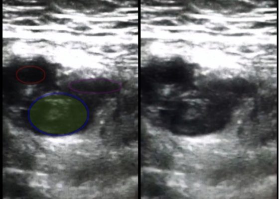
As shown in the still image of the performed ultrasound, a transverse view of the proximal-thigh revealed a visible thrombus (green shading) occluding the lumen of the left common femoral vein (blue ring), which was non-compressible when direct pressure was applied to the probe. Also visible is a patent and compressible branch of the common femoral vein (purple ring) and the femoral artery (red ring), highlighted by its thick vessel wall and pulsatile motion.
UltrasoundCardiology/VascularVisual EM
Creative Commons images

Bedside Echocardiography for Rapid Diagnosis of Malignant Cardiac Tamponade

Alaina Brinley, MD, PhD*, Maili Alvarado, MD* and J Christian Fox, MD*

DOI: https://doi.org/10.21980/J82S38Issue 2:1[mrp_rating_result]
The video shows a subxiphoid view of the heart with evidence of a large pericardial effusion with tamponade – note the anechoic stripe in the pericardial sac (see red arrow). This video demonstrates paradoxical right ventricular collapse during diastole and right atrial collapse during systole which is indicative of tamponade.1,2 Figure 1 is from the same patient and shows sonographic pulsus paradoxus. This is an apical 4 chamber view of the heart with the sampling gate of the pulsed wave doppler placed over the mitral valve. The Vpeak max and Vpeak min are indicated. If there is more than a 25% difference with inspiration between these 2 values, this is highly suggestive of tamponade.1 In this case, there is a 32.4% difference between the Vpeak max 69.55 cm/s and Vpeak min 46.99 cm/s.
Cardiology/VascularHematology/OncologyUltrasoundVisual EM
Creative Commons images

Takotsubo (Stress) Cardiomyopathy

Justin J Hourmozdi, MD*, Julian P Suszanski, MD* and Jacqueline M Pflaum, MD*^

DOI: https://doi.org/10.21980/J8Z309Issue 2:1[mrp_rating_result]
Bedside echocardiography showed the findings consistent with Takotsubo cardiomyopathy. Echocardiographic images are shown in subxiphoid (A) and apical four chamber (B) views. Note the apical ballooning appearance (asterisk) of the left ventricle (LV).
Cardiology/VascularVisual EM
Creative Commons images

Intraocular Foreign Body: Ultrasound and CT Findings

Maja Feldman, MPP*, Kristi Shigyo, MD^ and Amy Kaji, MD^

DOI: https://doi.org/10.21980/J8JS3MIssue 2:1[mrp_rating_result]
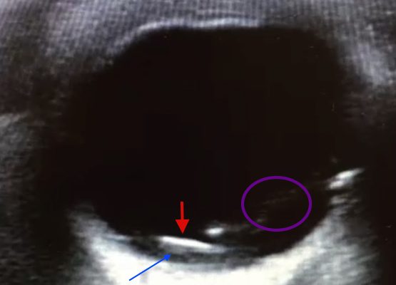
Point of care ultrasound revealed a mobile, radiolucent hyperechoic structure (see red arrow) with reverberation within the posterior chamber (see blue arrow), likely a metallic foreign body. Linear areas of mobile hyperechoic material revealed possible vitreous hemorrhage (see purple circular area). Orbital non-contrast CT confirmed a 3 mm metallic focus within the dependent portion of the left globe, lodged in the posterior sclera, with some vitreous hemorrhage but no evidence of globe rupture. Ophthalmology was consulted and the patient was taken to surgery later that night.
OphthalmologyVisual EM
Creative Commons images

Cholelithiasis: WES Sign

Hamid Ehsani-Nia, MS* and Shannon Toohey, MD, MAEd^

DOI: https://doi.org/10.21980/J8X300Issue 2:1[mrp_rating_result]
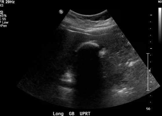
Abdominal ultrasound showed the classic presentation of the Wall-Echo-Shadow (WES) sign. The superficial aspect of the gallbladder wall is represented by a hyperechogenic curve. Below this, bile fluid is represented by hypoechogenicity. Underneath the bile fluid is the echo of the dense border created by the collection of gallstones, represented by a hyperechogenic curve. Due to the high density of the gallstones, nothing deeper can be visualized (including other gallstones or the far end of the gallbladder); this is the shadow.
Abdominal/GastroenterologyUltrasoundVisual EM
Creative Commons images

FAST Exam: Normal Suprapubic

[mrp_rating_result]
  Keywords: radiology, trauma, normal, suprapubic, FAST exam, videos, ultrasound, US
Trauma
Creative Commons images

FAST Exam: Normal Morrison’s Pouch

[mrp_rating_result]
Keywords: radiology, normal, renal, Morrison’s pouch, trauma, liver, ultrasound, video, FAST exam
Trauma
Creative Commons images

FAST Exam: Normal Morrison’s Pouch

[mrp_rating_result]
  Keywords: radiology, normal, renal, Morrison’s pouch, trauma, liver, ultrasound, FAST exam
Trauma
«‹10›
Page 10 of 11

JETem is an online, open access, peer-reviewed, journal-repository for EM educators. We are PMC Indexed.

Most Viewed

  • Simple Auricular Hematoma (“Cauliflower Ear”) Task Trainer
  • The Three Breath Method: Teaching Coping Mechanisms for Processing Patient Death
  • Clinical Decision Making: Acute Ischemic Stroke with Large Vessel Occlusion
  • PRIMER: Premedical Introduction to Mentored Emergency Research: Connecting Undergraduate Assistants and Emergency Medicine Residents in a Community Hospital Setting
  • Dangerous Dysrhythmias in the ED: An Interactive EKG Workshop for Emergency Medicine Clerkship Learners

Visit Our Collaborators


Creative Commons Licence
This work is licensed under a Creative Commons Attribution 4.0 International License.

About

Education

Learners should benefit from active learning. JETem accepts submissions of team-based learning, small group learning, simulation, podcasts, lectures, innovations, curricula, question sets, and visualEM.

Scholarship

We believe educators should advance through the scholarship of their educational work. JETem gives educators the opportunity to publish scholarly academic work so that it may be widely distributed, thereby increasing the significance of their results.

Links

  • Home
  • Aim and Scope
  • Current Issue
  • For Reviewers
  • Instructions for Authors
  • Contact Us

Newsletter

Sign up to receive updates from JETem regarding newly published issues and findings.

Copyright Creative Commons Attribution 4.0 International