JETem
  • Home
  • About
    • Aim and Scope
    • Our Team
    • Editorial Board
    • FAQ
  • Issues
    • Current Issue
    • Ahead of Print
    • Past Issues
  • Visual EM
    • Latest Visual EM
    • Search Visual EM
    • Thumbnail Library
  • For Authors
    • Instructions for Authors
    • Submit to JETem
    • Photo Consent
    • Policies
      • Peer Review Policy
      • Copyright Policy
      • Editorial Policy, Ethics and Responsibilities
      • Conflicts of Interest & Informed Consent
      • Open Access Policy
      • Acceptance Fee
  • For Reviewers
    • Instructions for JETem Reviewers
    • Interested in Being a JETem Reviewer?
  • Topic
    • Abdominal/Gastroenterology
    • Administration
    • Board Review
    • Cardiology/Vascular
    • Certifying Exam Cases
      • Clinical Decision-Making
      • Prioritization
      • Communication
      • Procedure & Ultrasound
    • Clinical Informatics, Telehealth and Technology
    • Dermatology
    • EMS
    • Endocrine
    • ENT
    • Faculty Development
    • Genitourinary
    • Geriatrics
    • Hematology/Oncology
    • Infectious Disease
    • Miscellaneous (stats, etc)
    • Neurology
    • Ob/Gyn
    • Ophthalmology
    • Orthopedics
    • Pediatrics
    • Pharmacology
    • Procedures
    • Psychiatry
    • Renal/Electrolytes/Nutrition
    • Respiratory
    • Social Determinants of Health
    • Toxicology
    • Trauma
    • Ultrasound
    • Urology
    • Wellness
    • Wilderness
  • Modality
    • Certifying Exam Practice Cases
      • Clinical Care Cases
      • Communication & Procedural Cases
      • Old School Oral Board Cases
    • Curricula
    • Innovations
    • Lectures
    • Podcasts
    • Simulation
    • Small Group Learning
    • Team Based Learning (TBL)
    • Visual EM
  • Contact Us

CT

Creative Commons images

Renal Infarction from Type B Aortic Dissection

Marit Tweet, MD* and James Roy Waymack, MD*

DOI: https://doi.org/10.21980/J8HG9G Issue 2:3[mrp_rating_result]
Initial abdominal images demonstrated a dissection flap; therefore, a CTA of the chest was also obtained. These images revealed a Stanford type B aortic dissection beginning just distal to the left subclavian artery and extending to the origin of the inferior mesenteric artery. The right renal artery arose from the true lumen of the dissection while the left renal artery arose from the false lumen. This case is interesting as imaging shows the lack of perfusion to the left kidney, residing in the retroperitoneum, which correlates with her non-descript abdominal and left flank pain.
Cardiology/VascularRenal/ElectrolytesVisual EM
Creative Commons images

Esophageal Perforation

Valentina Park, BS* and Jason Mefford, MD^

DOI: https://doi.org/10.21980/J8K91B Issue 2:3[mrp_rating_result]
History of present illness: A 51-year-old male with history of gastroesophageal reflux disease status post multiple endoscopies presented to the emergency department with severe abdominal pain. Paramedics reported the patient appeared diaphoretic on arrival and maintained stable vital signs during transit. The patient reported taking Prilosec that morning before eating breakfast, after which he felt like something was stuck in
Abdominal/GastroenterologyVisual EM
Creative Commons images

Globe Rupture

Reid Honda, MD*

DOI: https://doi.org/10.21980/J8N91Z Issue 2:3[mrp_rating_result]
The patient’s computed tomography (CT) head demonstrated a deformed left globe, concerning for ruptured globe. The patient had hyperdense material in the posterior segment (see green arrow), consistent with vitreous hemorrhage. CT findings that are consistent with globe rupture may include a collapsed globe, intraocular air, or foreign bodies.
OphthalmologyVisual EM
Creative Commons images

Ectopic Kidney

John Costumbrado, MD, MPH*, Reid Honda, MD^ and Eric McCoy, MD, MPH^

DOI: https://doi.org/10.21980/J89058 Issue 2:3[mrp_rating_result]
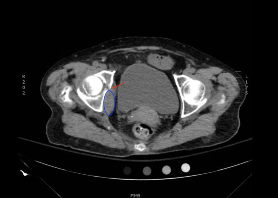
CT of the abdomen and pelvis revealed a normal left kidney and an ectopic, malrotated right kidney located in the pelvis (see white arrow).
Renal/ElectrolytesVisual EM
Creative Commons images

Ruptured Abdominal Aortic Aneurysm

Jessica Andrusaitis, BS, MS* and Jonathan Peña, MD*

DOI: https://doi.org/10.21980/J8FP6SIssue 2:3[mrp_rating_result]
CTA demonstrated a ruptured 7.4 cm infrarenal abdominal aortic aneurysm with a large left retroperitoneal hematoma.
Cardiology/VascularVisual EM
Creative Commons images

Open Pneumothorax

Bart Paull, MD*, Zachary Jarou, MD* and David Richards, MD*

DOI: https://doi.org/10.21980/J88036 Issue 2:3[mrp_rating_result]
A large chest wound was clinically obvious. A chest radiograph performed after intubation showed subcutaneous emphysema, an anterior rib fracture, and a right-sided pneumothorax. He was then taken to the operating room for further management.
TraumaRespiratoryVisual EM
Creative Commons images

Dense MCA Sign

Richard J Chen, MD* and Grant Wei, MD*

DOI: https://doi.org/10.21980/J8CS66Issue 2:3[mrp_rating_result]
A non-contrast computed tomography (CT) scan showed a hyperdensity along the right middle cerebral artery (MCA) consistent with acute thrombus. The red arrow highlights the hyperdensity in the annotated image.
NeurologyVisual EM
Creative Commons images

Acute, massive pulmonary embolism with right heart strain and hypoxia requiring emergent tissue plasminogen activator (TPA) infusion

Jonathan Patane, MD* and Wirachin Hoonpongsimanont, MD*

DOI: https://doi.org/10.21980/J84K5K Issue 2:2[mrp_rating_result]
CT angiogram showed multiple large acute pulmonary emboli, most significantly in the distal right main pulmonary artery (image 1 and 2). Additional pulmonary emboli were noted in the bilateral lobar, segmental, and subsegmental levels of all lobes. There was a peripheral, wedge-shaped consolidation surrounded by groundglass changes in the posterolateral basal right lower lobe that was consistent with a small lung infarction (image 3).
Cardiology/VascularRespiratoryVisual EM
«‹15›»
Page 15 of 17

JETem is an online, open access, peer-reviewed, journal-repository for EM educators. We are PMC Indexed.

Most Viewed

  • Escape Intern Orientation! — A Capstone and Team Building Activity for New EM Interns
  • Enhancing Relationship-Centered Communication and Feedback in Emergency Medicine Through Applied Improvisation (EM-PROV)
  • Postpartum Complications Modified TBL
  • Midline Catheters: A Novel Curriculum for Emergency Medicine Residents
  • Stopping Fistula Hemorrhage without Bleeding Time and Money – A Low Cost, Low Resource Hemodialysis Fistula Model for Emergency Medicine Residents

Visit Our Collaborators


Creative Commons Licence
This work is licensed under a Creative Commons Attribution 4.0 International License.

About

Education

Learners should benefit from active learning. JETem accepts submissions of team-based learning, small group learning, simulation, podcasts, lectures, innovations, curricula, question sets, and visualEM.

Scholarship

We believe educators should advance through the scholarship of their educational work. JETem gives educators the opportunity to publish scholarly academic work so that it may be widely distributed, thereby increasing the significance of their results.

Links

  • Home
  • Aim and Scope
  • Current Issue
  • For Reviewers
  • Instructions for Authors
  • Contact Us

Newsletter

Sign up to receive updates from JETem regarding newly published issues and findings.

Copyright Creative Commons Attribution 4.0 International