JETem
  • Home
  • About
    • Aim and Scope
    • Our Team
    • Editorial Board
    • FAQ
  • Issues
    • Current Issue
    • Ahead of Print
    • Past Issues
  • Visual EM
    • Latest Visual EM
    • Search Visual EM
    • Thumbnail Library
  • For Authors
    • Instructions for Authors
    • Submit to JETem
    • Photo Consent
    • Policies
      • Peer Review Policy
      • Copyright Policy
      • Editorial Policy, Ethics and Responsibilities
      • Conflicts of Interest & Informed Consent
      • Open Access Policy
      • Acceptance Fee
  • For Reviewers
    • Instructions for JETem Reviewers
    • Interested in Being a JETem Reviewer?
  • Topic
    • Abdominal/Gastroenterology
    • Administration
    • Board Review
    • Cardiology/Vascular
    • Certifying Exam Cases
      • Clinical Decision-Making
      • Prioritization
      • Communication
      • Procedure & Ultrasound
    • Clinical Informatics, Telehealth and Technology
    • Dermatology
    • EMS
    • Endocrine
    • ENT
    • Faculty Development
    • Genitourinary
    • Geriatrics
    • Hematology/Oncology
    • Infectious Disease
    • Miscellaneous (stats, etc)
    • Neurology
    • Ob/Gyn
    • Ophthalmology
    • Orthopedics
    • Pediatrics
    • Pharmacology
    • Procedures
    • Psychiatry
    • Renal/Electrolytes/Nutrition
    • Respiratory
    • Social Determinants of Health
    • Toxicology
    • Trauma
    • Ultrasound
    • Urology
    • Wellness
    • Wilderness
  • Modality
    • Certifying Exam Practice Cases
      • Clinical Care Cases
      • Communication & Procedural Cases
      • Old School Oral Board Cases
    • Curricula
    • Innovations
    • Lectures
    • Podcasts
    • Simulation
    • Small Group Learning
    • Team Based Learning (TBL)
    • Visual EM
  • Contact Us

CT

Creative Commons images

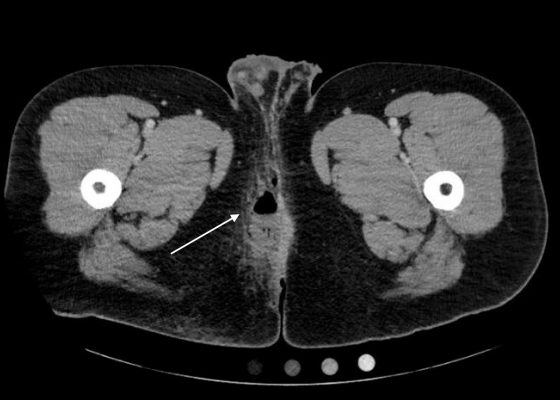
Perianal Abscess

Luke Walls-Smith, BS* and Jonathan Peña, MD*

DOI: https://doi.org/10.21980/J8QP81Issue 3:1[mrp_rating_result]
Computed Tomography (CT) of the Pelvis with intravenous (IV) contrast revealed a 5.7 cm x 2.4 cm air-fluid collection in the right perianal soft tissue along the right gluteal cleft, with surrounding fat stranding, consistent with a perianal abscess with cellulitis.
Abdominal/GastroenterologyInfectious DiseaseVisual EM
Creative Commons images

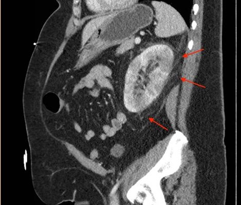
Acute Pyelonephritis with Perinephric Stranding on CT

Ronald Goubert* and Jonathan Peña, MD*

DOI: https://doi.org/10.21980/J8BH0VIssue 3:1[mrp_rating_result]
A CT abdomen and pelvis with IV contrast showed neither nephrolithiasis nor diverticulitis, and instead showed heterogeneous enhancement of the left kidney with mild edematous enlargement and striated left nephrogram. Significant perinephric stranding (red arrows) was also noted and was consistent with severe acute pyelonephritis.
Renal/ElectrolytesInfectious DiseaseVisual EM
Creative Commons images

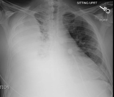
Empyema

Jonathan Patane, MD* and Daniel Ritter, BS^

DOI: https://doi.org/10.21980/J86P9RIssue 3:1[mrp_rating_result]
The chest X-ray shows a large fluid collection in the right lung demonstrated by the opacification that blunts the costophrenic angle on the right side. There is also a meniscus present, which is generally indicative of fluid.  Chest computed tomography (CT) demonstrated an infiltrate with a mixture of densities within the same collection, consistent with a loculated effusion and concerning for an empyema.
RespiratoryInfectious DiseaseVisual EM
Creative Commons images

Oropharynx Ulceration

Lauren Sylwanowicz, MD* and Andrew Wong, MD, MBA*

DOI: https://doi.org/10.21980/J87W60 Issue 2:4[mrp_rating_result]
The photograph demonstrates an area of ulcerative tissue at the left palatine tonsil without surrounding erythema or purulent drainage. The computed tomography (CT) scan shows a large ulceration of the left soft palate and palatine tonsil (red arrow). There is no evidence of skull base osteomyelitis. There is suppurative lymphadenopathy with partial left jugular vein compression due to mass effect (yellow highlight). There is mild nasopharyngeal airway narrowing with architectural distortion (blue arrow), but no other evidence of airway obstruction. 
ENTVisual EM
Creative Commons images

An Elderly Male with Amyand’s Hernia

Saema Said, MD* and Roozbeh Houshyar, MD^

DOI: https://doi.org/10.21980/J80D13 Issue 2:4[mrp_rating_result]
Ultrasound of the right scrotum shows a right inguinal hernia with an air-containing loop of bowel (white arrow) and a non-compressible appendix (yellow arrow). Coronal and axial views of abdomen-pelvis CT show a right inguinal hernia containing a loop of small bowel (white arrow) and appendix (yellow arrow).
Abdominal/GastroenterologyVisual EM
Creative Commons images

Acetabular Fracture

Chad Correa* and Sari Lahham, MD^

DOI: https://doi.org/10.21980/J8BK8K Issue 2:4[mrp_rating_result]
The non-contrast CT images show a minimally displaced comminuted fracture of the right acetabulum involving the acetabular roof, medial and anterior walls (red arrows), with associated obturator muscle hematoma (blue oval).
OrthopedicsVisual EM
Creative Commons images

Choledocholithiasis

Jonathan Peña, MD*

DOI: https://doi.org/10.21980/J8Q62X Issue 2:3[mrp_rating_result]
Computed tomography (CT) was significant for two large gallstones measuring 1.1 centimeters impacted at the level of the pancreatic head with associated common bile duct (CBD) dilatation.
Abdominal/GastroenterologyVisual EM
Creative Commons images

Volvulus

Sari Lahham, MD, MBA*, Kathryn Bennett, BS* and Mohammad Helmy, MD†

DOI: https://doi.org/10.21980/J8JH0Q Issue 2:3[mrp_rating_result]
Upright and supine frontal radiographs of the abdomen demonstrate gas dilation of the large bowel from the level of the cecum to the sigmoid colon with air fluid levels (yellow arrows). There is a swirled configuration of the distal descending to sigmoid colon indicating the level of the volvulus (dashed yellow line) and giving rise to the classic “coffee bean” sign (dotted white tracing). Note the elevated left hemidiaphragm on the upright view reflecting abdominal distention with increased intra-abdominal pressure (red arrow).
Abdominal/GastroenterologyVisual EM
«‹14›»
Page 14 of 17

JETem is an online, open access, peer-reviewed, journal-repository for EM educators. We are PMC Indexed.

Most Viewed

  • Escape Intern Orientation! — A Capstone and Team Building Activity for New EM Interns
  • Enhancing Relationship-Centered Communication and Feedback in Emergency Medicine Through Applied Improvisation (EM-PROV)
  • Postpartum Complications Modified TBL
  • Midline Catheters: A Novel Curriculum for Emergency Medicine Residents
  • Stopping Fistula Hemorrhage without Bleeding Time and Money – A Low Cost, Low Resource Hemodialysis Fistula Model for Emergency Medicine Residents

Visit Our Collaborators


Creative Commons Licence
This work is licensed under a Creative Commons Attribution 4.0 International License.

About

Education

Learners should benefit from active learning. JETem accepts submissions of team-based learning, small group learning, simulation, podcasts, lectures, innovations, curricula, question sets, and visualEM.

Scholarship

We believe educators should advance through the scholarship of their educational work. JETem gives educators the opportunity to publish scholarly academic work so that it may be widely distributed, thereby increasing the significance of their results.

Links

  • Home
  • Aim and Scope
  • Current Issue
  • For Reviewers
  • Instructions for Authors
  • Contact Us

Newsletter

Sign up to receive updates from JETem regarding newly published issues and findings.

Copyright Creative Commons Attribution 4.0 International