
Visual EM

A Case Report of Corneoscleral Laceration with Open Globe Injury and Iris Prolapse
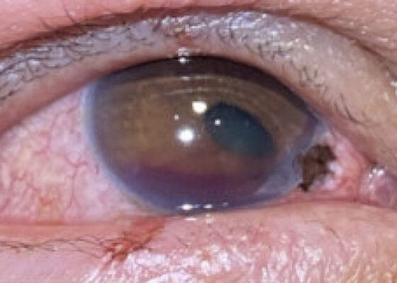
DOI: https://doi.org/10.5070/M5.52326Closer examination of the right eye revealed a 2 mm x 1 mm corneoscleral laceration to the 4-5 o'clock position at the border of zone I (yellow highlighting) and II (blue highlighting) with iris prolapse (yellow star) and plugging. Additionally, the affected eye revealed a peaked pupil (cyan highlighting) in the inferonasal direction with associated corectopia and a 2 mm Grade I hyphema (pink highlighting). Immediately inferior to the corneal laceration, 1 mm epithelial injury of the lower palpebra was present (green cross). Fluorescein exam demonstrated uptake at the 4-5 o'clock position (yellow arrow) but was negative for Seidel’s sign.
Diagnosis of Sinonasal Carcinoma in the Emergency Department: A Case Report Highlighting Red Flag Symptoms
DOI: https://doi.org/10.5070/M5.52257Physical exam revealed a gross deformity of the left side of the face with soft tissue swelling with no overlying skin changes. The left eye was proptotic and completely immobile, with cranial nerve (CN) 3, 4, and 6 palsies. Additionally, a large obstructive mass was noted in the left naris with resultant rightward displacement of the nasal septum. Intraocular pressures (IOPs) were measured to be 6 mmHg in the right eye and 11 mmHg in the left eye.
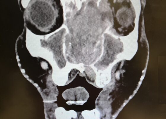
Imaging, including maxillofacial and neck soft tissue CTs revealed a large, destructive, soft tissue mass centered in the nasal cavity with significant osseous destruction of the midface and skull base (red highlighted area). There was mild intracranial extension with the mass abutting or infiltrating the inferior frontal lobes (green highlighted area). The nasal cavity and nasopharynx were obstructed (orange highlighted area). The mass invaded the left orbit with associated left-sided proptosis, left globe deformity, and compressed the left optic nerve and the left optic chiasm (blue arrows).
Trapped In Transit – A Case Report of a Pediatric Gastric Bezoar Causing Intermittent Small Bowel Obstruction
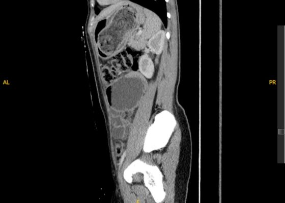
DOI: https://doi.org/10.5070/M5.52259The CT scan identified a heterogenous, mass-like lesion extending from the gastric fundus to the antrum, along with a small bowel obstruction (SBO) and significant small bowel dilation. An additional smaller heterogenous mass-like lesion was seen in the small bowel, causing the SBO. Given the mass-like lesion’s mixed attenuation and presence of internal air, a gastric bezoar was strongly suspected.
Overlooked and Undernourished: A Case Report of Scurvy Linked to Food Insecurity
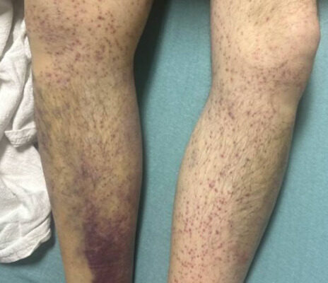
DOI: https://doi.org/10.5070/M5.52313Images A and B of the lower extremities show what initially appeared to be a petechial rash with scattered ecchymoses bilaterally, primarily on the right lower leg. On closer examination, images C and D more clearly show these spots to be flat and non-blanching, indicative of follicular hyperkeratosis with perifollicular hemorrhages. The classic coiled or corkscrew hair findings are not seen in these images. Image E shows evidence of palatal ecchymosis, without evidence of gingival bleeding, likely due to loss of dentition. This constellation of exam findings reflects the production of weakened or dysfunctional collagen in connective tissue and vascular structures and is strongly suggestive of the clinical diagnosis of scurvy. Image F shows resolution of the patient’s dermatologic findings after three weeks of treatment.
A Case Report of a 36-year-old Male Diagnosed with a Spontaneous Coronary Artery Dissection
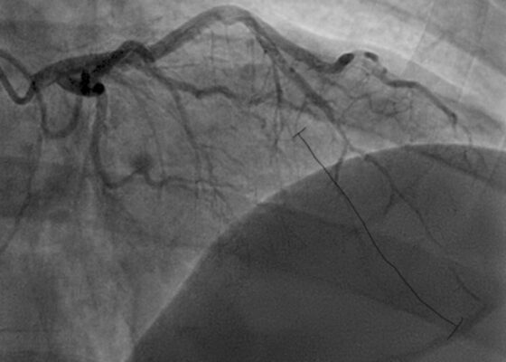
DOI: https://doi.org/10.5070/M5.52022The initial ECG obtained from the patient shows subtle ST-segment elevation noted in leads I, aVL, and V2-V5, suggestive of pathology of the left anterior descending artery. The results of the catheterization revealed a spontaneous coronary artery dissection of the distal portion of the left anterior descending coronary artery, which can be seen in the image of the angiogram, with the diseased portion notated between the brackets.
A Case Report of an Atypical Presentation of Fournier’s Gangrene
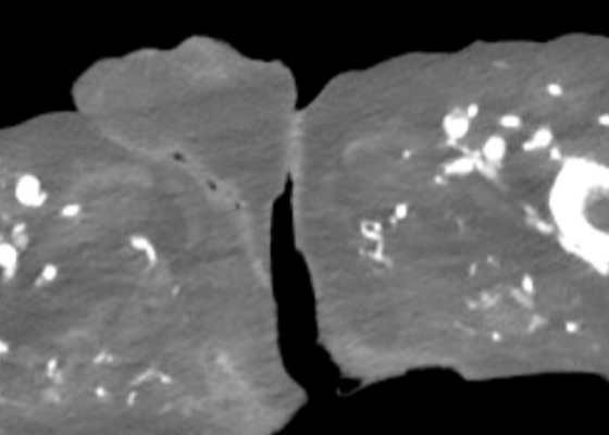
DOI: https://doi.org/10.5070/M5.52203A computed tomography (CT) scan of the abdomen and pelvis was significant for scrotal fluid and punctate gas locules (red arrow) without discrete evidence of invasion into the adjacent soft tissues, suspicious for Fournier’s gangrene. There was also fluid collection centered around the seminal vesicles suggestive of an abscess.
Open Chest Wound with Sternal Fracture in the Emergency Department, a Case Report
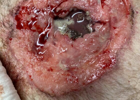
DOI: https://doi.org/10.5070/M5.52202The image demonstrates the large chronic-appearing wound of the patient’s anterior chest as well as the visible fractured segments of the patient’s exposed sternum. The sternum is necrotic appearing concerning for a chronic process including osteomyelitis and malignancy. Purulent drainage is visible on the wound consistent with an infectious process.
Effects of Volume Overload: A Case Report of an Edema Bulla
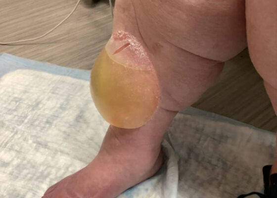
DOI: https://doi.org/10.5070/M5.52206This image shows a large edema bulla on the patient's right shin. The bulla is 10 x 10 cm, filled with serous fluid, has a spontaneously occurring defect in the skin of the superior portion of the bulla, and is non-erythematous. The bulla is much larger than the 1-5 cm edema bullae described in the literature. As edema bulla is primarily a clinical diagnosis, taking the full history and physical exam into account is essential to recognize these bullae.