
Orthopedics
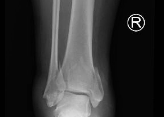

Trimalleolar Fracture
DOI: https://doi.org/10.21980/J8PP46Anteroposterior (AP), lateral, and oblique x-ray views were obtained. The AP view revealed a displaced spiral fracture of the lateral malleolus (red) and a vertical fracture of the medial malleolus (blue). The lateral view revealed a displaced fracture of the posterior malleolus, tibial plafond (yellow), and the oblique view showed widening of the distal tibiofibular syndesmosis (green). CT scans confirmed trimalleolar fracture, annotated with the same colored lines (red, lateral malleolus; blue, medial malleolus; yellow, posterior malleolus).

Perilunate Dislocation
DOI: https://doi.org/10.21980/J8F59RIn the left lateral wrist x-ray, the lunate is dislocated from the rest of the wrist bones but still articulates with the radius. The capitate does not sit within the distal articulation of the lunate and is displaced dorsally. Additionally, a line drawn through the radius and lunate fails to intersect with the capitate. This is consistent with a perilunate dislocation. This is compared to a lunate dislocation, where the lunate itself is displaced and turned ventrally (spilled teacup) and the proximal aspect does not articulate with the radius.

Femoral Neck Fracture
DOI: https://doi.org/10.21980/J89G6GIn the anteroposterior view bilateral hip x-ray, there is an evident loss of Shenton’s line on the left when compared to the normal right, indicative of a fracture in the left femoral neck. This correlates with findings seen on pelvic CT, which reveals both a subcapital fracture and transcervical fracture. The neck of the femur is displaced superiorly relative to the head of the femur while the head of the femur remains in its anatomical position within the acetabulum.

Posterior Elbow Dislocation
DOI: https://doi.org/10.21980/J8X593Elbow dislocations are classified by the position of the radio-ulnar joint relative to the humerus.1 Images 1, 2, and 3 show a left posterior elbow dislocation; the radius and ulna (red lines) are displaced posteriorly with respect to the distal humerus (blue line). The lateral view of the elbow most clearly shows this: trochlear notch of the ulna (red line) is empty and displaced posteriorly relative to the trochlea (blue line). There is no associated fracture. Images 4 and 5 show the elbow status-post reduction, demonstrating proper alignment of the distal humerus (blue line) with the radius and ulna (red lines).
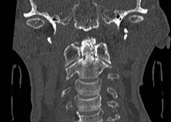
Odontoid Fracture: Computed Tomography
DOI: https://doi.org/10.21980/J8NP4WComputed Tomography (CT) of the cervical spine showed a stable, acute, non-displaced fracture of the odontoid process extending into the body of C2, consistent with a Type III Odontoid Fracture. He was evaluated by orthopedic spine service who recommended conservative, non-operative management.

Open Dislocation of Fifth Digit
DOI: https://doi.org/10.21980/J8J01XPhysical exam revealed an open dislocation of the proximal interphalangeal joint (PIP) of the right fifth digit. X-ray confirmed dislocation and revealed no fractures. The patient received a tetanus booster, Cefazolin, and the dislocation was then washed out and reduced. Multiple reduction attempts were made and were only successful once the metacarpophalangeal joints were held in 90 degree flexion, which relaxed the lateral bands and enabled the finger to be reduced.

Morel-Lavallée Lesion
DOI: https://doi.org/10.21980/J88G65On physical examination, the patient was noted to have a nearly “watermelon-sized” fluctuant mass to his right lateral superior quadriceps with multiple overlying abrasions (Image 1). Computed tomography (CT) scans of the area showed a large heterogeneous collection measuring roughly 37x9.5x16 centimeters in the subcutaneous adipose layer of the lateral right thigh (Image 2), while ultrasonography revealed a complex fluid collection containing some nodular solid components and debris (Image 3). Additionally, radiographs confirmed multiple fractures including most significantly a pelvic ring fracture. Surgical debridement, evacuation, and sclerodhesis were performed nine weeks post injury to allow overlying abrasions to heal prior to intervention.
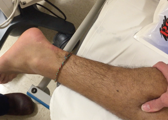
Thompson Test in Achilles Tendon Rupture
DOI: https://doi.org/10.21980/J8VC7SThe left Achilles tendon had a defect on palpation, while the right Achilles tendon was intact. When squeezing the right (unaffected) calf, the ankle spontaneously plantar flexed, indicating a negative (normal) Thompson test. Upon squeeze of the left (affected) calf, the ankle did not plantar flex, signifying a positive (abnormal) Thompson test. The diagnosis of left Achilles tendon rupture was confirmed intraoperatively one week later.