A-Lines below the Diaphragm – A Case Report of Occult Pneumoperitoneum
Correspondence should be addressed to Yong Hoon Lee, MD at nleeyh@gmail.com
DOI: https://doi.org/10.5070/M5.52389
ABSTRACT:
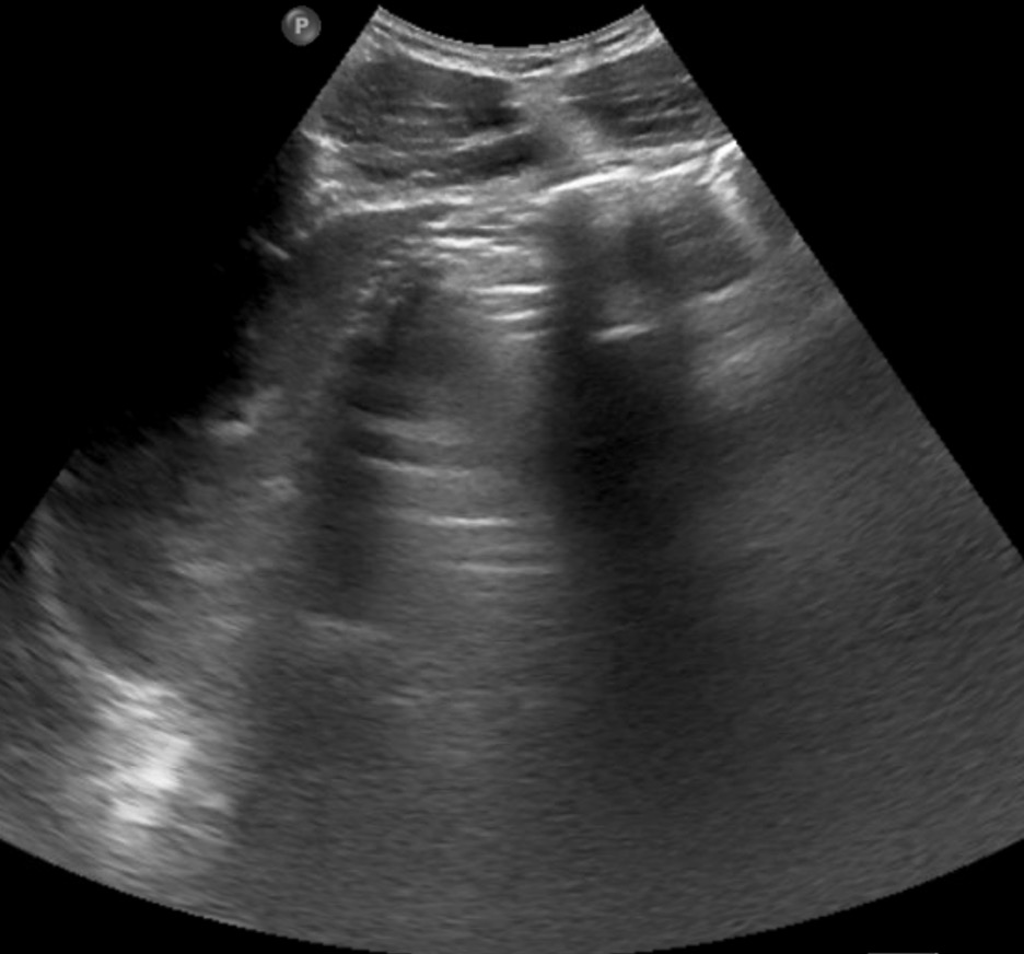
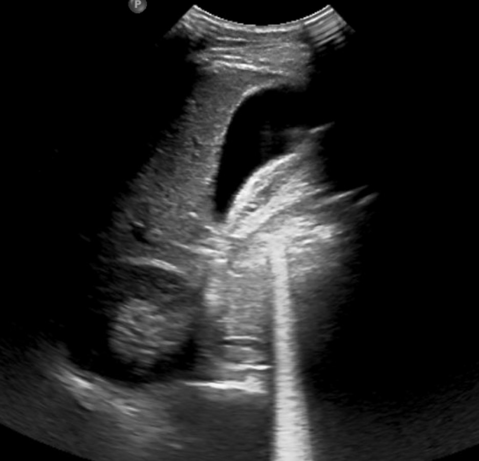
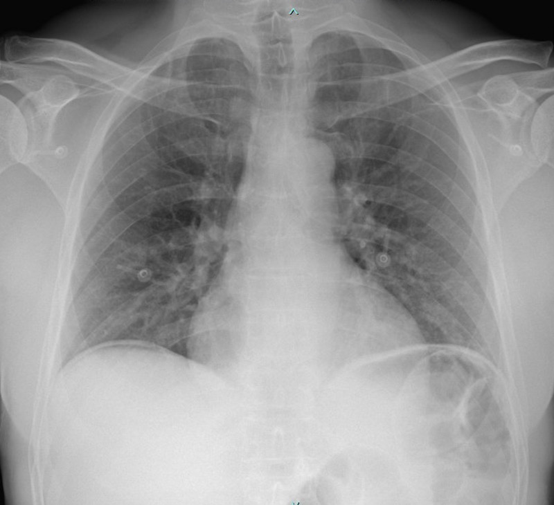
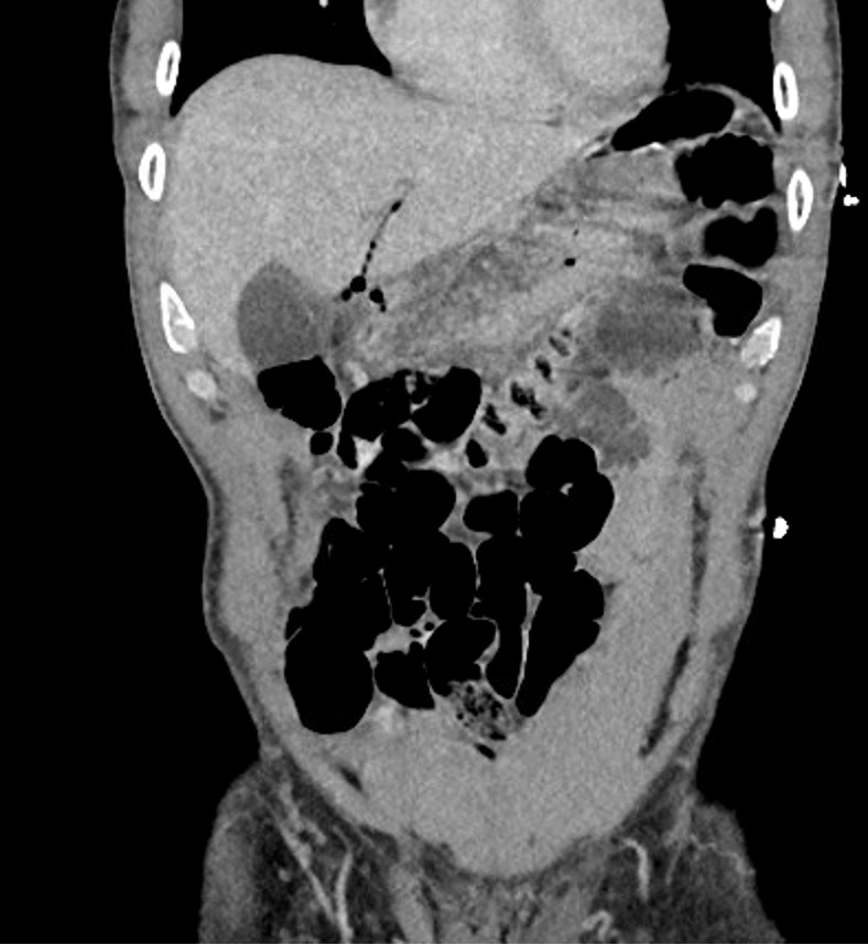
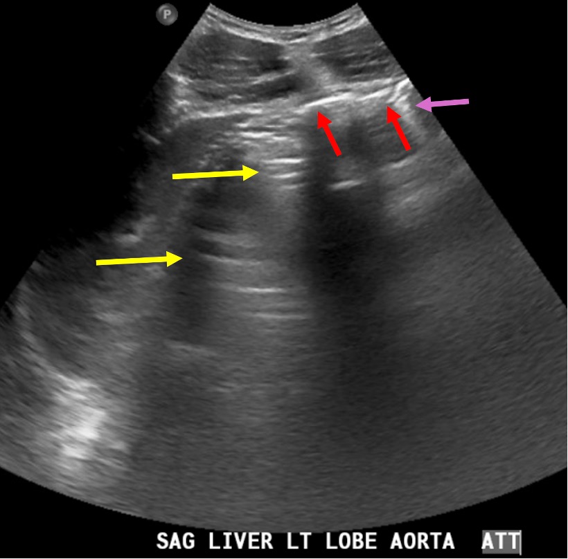
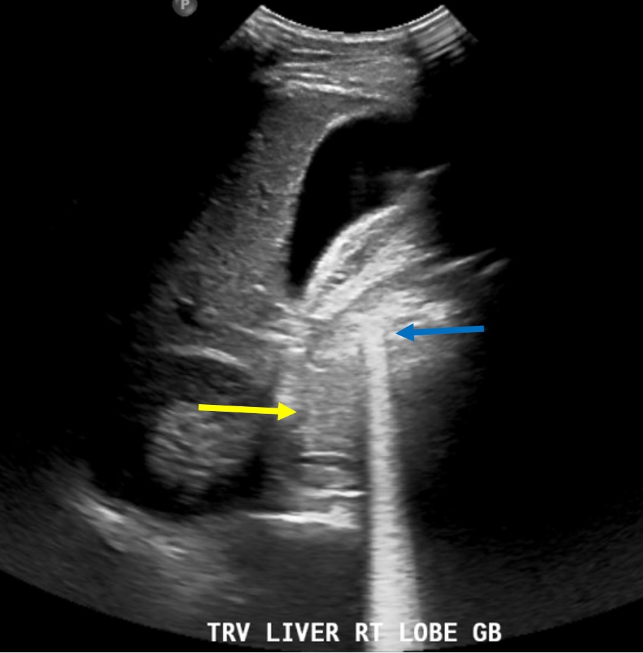
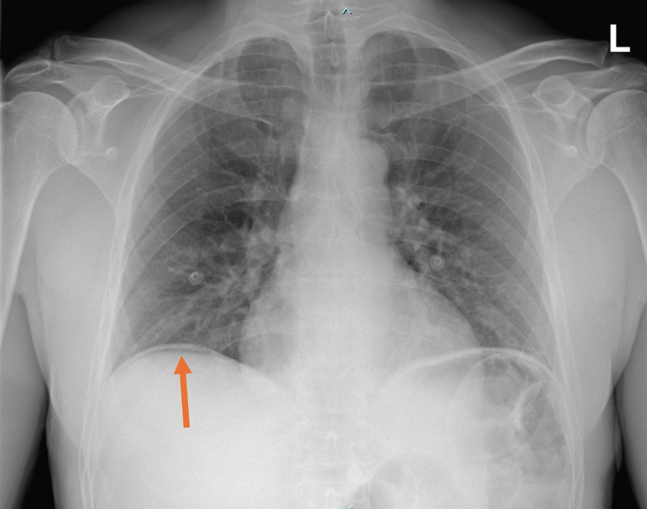
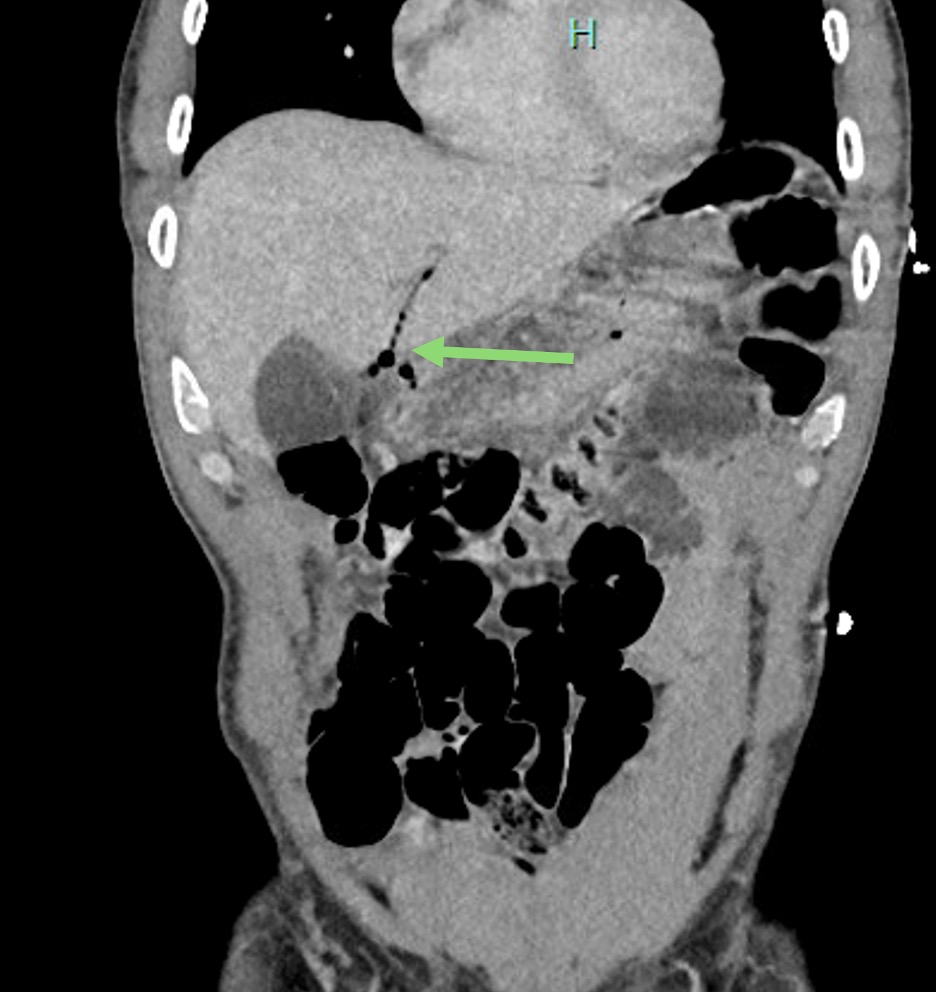
This case report highlights the rapid and sensitive diagnostic capabilities of a right upper quadrant (RUQ) ultrasound to diagnose pneumoperitoneum secondary to a hollow viscus rupture. The patient was a 55-year-old male with sudden onset epigastric and right upper quadrant pain. Symptoms were concerning for either acute cholecystitis and gastric ulcer or perforation. Immediate point of care ultrasound (POCUS) of the RUQ showed no signs of acute cholecystitis but showed concerning findings of intraabdominal free air given the enhanced peritoneal stripe sign, reverberation, and ring down artifacts. The radiologist did not note any signs of free air on the interpretation of the formal ultrasound. Eventually, the chest X-ray and computed tomography (CT) of the abdomen and pelvis showed air under the right diaphragm, fluid in the porta hepatis, and multiple foci of pneumoperitoneum. Broad spectrum antibiotics and fluid resuscitation were given. General surgery performed an emergency diagnostic laparotomy with repair of duodenal ulcer perforation. Upper gastrointestinal (GI) study on post-operative day three showed no complications, and patient has been doing well in follow-up clinic visits. In this case report, immediate recognition of signs of pneumoperitoneum would have expedited the treatment and management of the duodenal perforation. Although our emergency department was able to perform a CT scan of the abdomen and pelvis and have it read within two hours, this may not be possible in all emergency departments. Therefore, the use of POCUS in the evaluation of abdominal pain can identify potentially disastrous time-dependent pathology. It would be beneficial for emergency physicians to learn the signs of pneumoperitoneum on ultrasound since this requires emergent surgery.
Topics: Hollow viscus perforation, ultrasound, pneumoperitoneum, point-of-care ultrasound (POCUS).