
JETem
  • Home
  • About
    • Aim and Scope
    • Our Team
    • Editorial Board
    • FAQ
  • Issues
    • Current Issue
    • Ahead of Print
    • Past Issues
  • Visual EM
    • Latest Visual EM
    • Search Visual EM
    • Thumbnail Library
  • For Authors
    • Instructions for Authors
    • Submit to JETem
    • Photo Consent
    • Policies
      • Peer Review Policy
      • Copyright Policy
      • Editorial Policy, Ethics and Responsibilities
      • Conflicts of Interest & Informed Consent
      • Open Access Policy
      • Acceptance Fee
  • For Reviewers
    • Instructions for JETem Reviewers
    • Interested in Being a JETem Reviewer?
  • Topic
    • Abdominal/Gastroenterology
    • Administration
    • Board Review
    • Cardiology/Vascular
    • Certifying Exam Cases
      • Clinical Decision-Making
      • Prioritization
      • Communication
      • Procedure & Ultrasound
    • Clinical Informatics, Telehealth and Technology
    • Dermatology
    • EMS
    • Endocrine
    • ENT
    • Faculty Development
    • Genitourinary
    • Geriatrics
    • Hematology/Oncology
    • Infectious Disease
    • Miscellaneous (stats, etc)
    • Neurology
    • Ob/Gyn
    • Ophthalmology
    • Orthopedics
    • Pediatrics
    • Pharmacology
    • Procedures
    • Psychiatry
    • Renal/Electrolytes/Nutrition
    • Respiratory
    • Social Determinants of Health
    • Toxicology
    • Trauma
    • Ultrasound
    • Urology
    • Wellness
    • Wilderness
  • Modality
    • Certifying Exam Practice Cases
      • Clinical Care Cases
      • Communication & Procedural Cases
      • Old School Oral Board Cases
    • Curricula
    • Innovations
    • Lectures
    • Podcasts
    • Simulation
    • Small Group Learning
    • Team Based Learning (TBL)
    • Visual EM
  • Contact Us

Posts by JETem

Creative Commons images

Bladder Diverticulum – A Case Report

Savannah Tan, BS* and Sangeeta Sakaria, MD, MPH, MST*

DOI: https://doi.org/10.21980/J8635C Issue 5:4[mrp_rating_result]
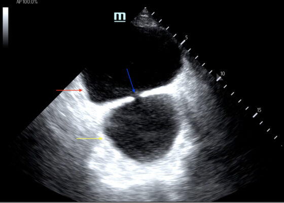
On examination, the patient was alert and oriented but in mild distress. Suprapubic fullness was noted upon abdominal palpation. Point of care ultrasound of the bladder showed two enlarged “bladders” with a central communication. Bedside total bladder volume was measured to be 1288 cm3 (the top “bladder” was measured to be 1011 cm3, while the bottom “diverticulum” was measured to be 277 cm3) by ultrasound. The POCUS stills of the patient’s bladder demonstrated the bladder (red arrow) and bladder diverticulum (yellow arrow) with a central communication (blue arrow) in the transverse and sagittal views.
Renal/ElectrolytesUltrasoundVisual EM
Creative Commons images

A Case Report of Ogilvie’s Syndrome in a 58-year-old Quadriplegic

Rosie Kumar, BS*, Brett Cowan, MD*, Daniel Quesada, MD* and Sage Wexner, MD*

DOI: https://doi.org/10.21980/J82922 Issue 5:4[mrp_rating_result]
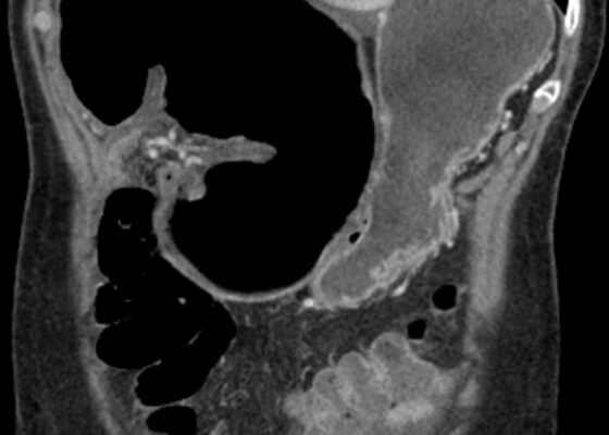
Plain radiograph of the patient's abdomen revealed a gaseous distention of the colon. This is demonstrated as noted in the abdominal x-ray as gaseous distention, most notably in the large bowel (arrows) including the rectal region (large circle). Follow up computed tomography (CT) scan affirmed severe pancolonic gaseous distention measuring up to 11.2 cm, compatible with colonic pseudo-obstruction as noted by the large red arrows. No anatomical lesion or mechanical obstruction was observed, as well as no evidence of malignancy or other acute process.
Abdominal/GastroenterologyVisual EM
Subtalar Dislocation Jetem

Cecal Volvulus Diagnosed with a Whirl Sign: A Case Report

Gregory K Sun, DO* and Brian Walsh, MD*

DOI: https://doi.org/10.21980/J8XM05 Issue 5:4[mrp_rating_result]
The CT image demonstrates a “whirl sign” (red arrow) which is indicative of a volvulus. This image occurs when bowel, mesentery and vasculature rotate around a transition point causing an image similar to a hurricane on a weather map. When seen on a CT scan, a whirl sign suggests a high likelihood of either a closed loop bowel obstruction or volvulus in the cecum, sigmoid or midgut. In any of the cases, seeing a whirl sign strongly increases the need for emergent surgical management.
Abdominal/GastroenterologyVisual EM
Creative Commons images

Paroxysmal Ventricular Standstill—A Case Report of all Ps and no QRS in Ventricular Asystole

Hamid Ehsani-Nia, DO* and Christopher Bryczkowski, MD*

DOI: https://doi.org/10.21980/J8SS79 Issue 5:4[mrp_rating_result]
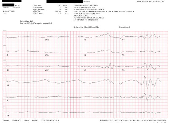
In route, it was proposed that this patient was suffering from a dysrhythmia due to the transient episodes of syncope with lack of ventricular activity on telemetry. Upon close examination of the rhythm strips as well as the ECG, P waves can be visualized without any accompanying QRS complexes lasting multiple seconds (ED ECG blue arrows). Additionally, the rhythm has an intrinsic rate of 100 beats per minute and has a consistent morphology with no evidence of ventricular activity due to the lack of QRS complexes. As a result, the rhythm likely originates in the atria with no passage of impulses into the ventricles through the atrioventricular (AV) node versus an accelerated ventricular rhythm where QRS complexes would be seen.8 These rhythm strips demonstrate an example of VS. There is preserved native atrial automaticity, with an intact sinoatrial (SA) node, with a complete lack of ventricular electrical activity
Cardiology/VascularVisual EM

Hemoptysis Due to Diffuse Alveolar Hemorrhage

Zoltan Buchwald, MD* and Amrita Vempati, MD*

DOI: https://doi.org/10.21980/J8ZP86 Issue 5:3[mrp_rating_result]
By the end of this simulation session, learners will be able to: (1) recognize worsening respiratory status of a patient with hemoptysis and intervene appropriately, (2) manage a patient with severe hemoptysis and perform appropriate ventilator management, (3) manage sinus tachycardia with QT prolongation on the ECG caused by cocaine and hypomagnesemia, (4) address various etiologies of hemoptysis, (5) discuss the causes of massive hemoptysis and management options, and (6) review ventilation strategies in an intubated hypoxic patient.
RespiratorySimulation

Improving Emergency Department Airway Preparedness in the Era of COVID-19: An Interprofessional, In Situ Simulation

Keiran J Warner, MD*, Ashley C Rider, MD*, James Marvel, MD*, Michael A Gisondi, MD*, Kimberly Schertzer, MD* and Kelly N Roszczynialski, MD, MS*

DOI: https://doi.org/10.21980/J8V06M Issue 5:3[mrp_rating_result]
At the conclusion of the simulation session, learners will be able to: 1) Understand the need to notify team members of a planned COVID intubation including: physician, respiratory therapist, pharmacist, nurse(s), and ED technician. 2) Distinguish between in-room and out-of-room personnel during high-risk aerosolizing procedures. 3) Distinguish between in-room and out-of-room equipment during high-risk aerosolizing procedures to minimize contamination. 4) Appropriately select oxygenation therapies and avoid high-risk aerosolizing procedures. 5) Manage high risk scenarios such as hypotension or failed intubation and be prepared to give push-dose vasoactive medications or place a rescue device such as an I-gel®.
Infectious DiseaseProceduresRespiratorySimulation

Eclampsia

Patrick G Meloy, MD*, Megan C Henn, MD*, Daniel Rutz, MD^ and Amit Bhambri, MD†

DOI: https://doi.org/10.21980/J8M93D Issue 5:3[mrp_rating_result]
At the end of this oral boards session, examinees will: 1) Demonstrate ability to obtain a complete medical history including a detailed obstetric history. 2) Demonstrate the ability to perform a detailed physical examination in a postpartum female patient who presents with a seizure. 3) Investigate the broad differential diagnoses which include electrolyte imbalances, brain tumor, meningitis or encephalitis, hemolysis, elevated liver enzymes, low platelets (HELLP) syndrome and eclampsia. 4) List the appropriate laboratory and imaging studies to differentiate eclampsia from other diagnoses (complete blood count, comprehensive metabolic panel, magnesium level, pregnancy testing, urinalysis, and computed tomography [CT] scan of the head). 5) Identify a postpartum eclampsia patient and manage appropriately (administer IV magnesium therapy, administer IV antihypertensive therapy, emergent consultation with an obstetrician). 6) Provide appropriate disposition to the intensive care unit after consulting with an obstetrician.
Ob/GynCertifying Exam Cases

Low Fidelity Trainer for Fiberoptic Scope Use in the Emergency Department

Garren Giles, DO*, Dominic Diprinzio, DO* and Jordana Haber, MD*

DOI: https://doi.org/10.21980/J8764B Issue 5:3[mrp_rating_result]
By the end of this training session, learners will be able to 1) list indications, contraindication, and complications in performing fiberoptic intubations, 2) know how to use and maneuver a fiberoptic scope, and 3) be able to successfully intubate the trainer model.
ProceduresInnovationsRespiratory
«‹39›»
Page 39 of 91

JETem is an online, open access, peer-reviewed, journal-repository for EM educators. We are PMC Indexed.

Most Viewed

  • Simple Auricular Hematoma (“Cauliflower Ear”) Task Trainer
  • The Three Breath Method: Teaching Coping Mechanisms for Processing Patient Death
  • Clinical Decision Making: Acute Ischemic Stroke with Large Vessel Occlusion
  • PRIMER: Premedical Introduction to Mentored Emergency Research: Connecting Undergraduate Assistants and Emergency Medicine Residents in a Community Hospital Setting
  • Dangerous Dysrhythmias in the ED: An Interactive EKG Workshop for Emergency Medicine Clerkship Learners

Visit Our Collaborators


Creative Commons Licence
This work is licensed under a Creative Commons Attribution 4.0 International License.

About

Education

Learners should benefit from active learning. JETem accepts submissions of team-based learning, small group learning, simulation, podcasts, lectures, innovations, curricula, question sets, and visualEM.

Scholarship

We believe educators should advance through the scholarship of their educational work. JETem gives educators the opportunity to publish scholarly academic work so that it may be widely distributed, thereby increasing the significance of their results.

Links

  • Home
  • Aim and Scope
  • Current Issue
  • For Reviewers
  • Instructions for Authors
  • Contact Us

Newsletter

Sign up to receive updates from JETem regarding newly published issues and findings.

Copyright Creative Commons Attribution 4.0 International