Issue 2:4
Erythema Migrans
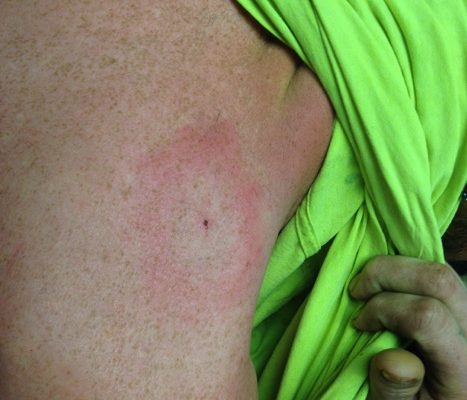
DOI: https://doi.org/10.21980/J8QW7QHistory of present illness: A 28-year-old male presented to the emergency department with a chief complaint of two weeks of headache, chills, and numbness in his hands. He reported removing a tick from his upper back approximately two weeks ago, but did not know how long the tick had been embedded. His review of symptoms was otherwise unremarkable. Significant findings:
Spontaneous Pneumothorax
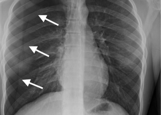
DOI: https://doi.org/10.21980/J8M33BInitial chest radiograph showed a 50% right-sided pneumothorax with no mediastinal shift, which can be identified by the sharp line representing the pleural lung edge (see arrows) and lack of peripheral lung markings extending to the chest wall. While difficult to accurately estimate volume from a two-dimensional image, a 2 cm pneumothorax seen on chest radiograph correlates to approximately 50% volume.1 The patient underwent insertion of a pigtail pleural drain on the right and repeat chest radiograph showed resolution of previously seen pneumothorax. Ultimately the pigtail drain was removed and chest radiograph showed clear lung fields without evidence of residual pneumothorax or pleural effusion.
Pediatric Esophageal Foreign Body
DOI: https://doi.org/10.21980/J8GD1FA radiopaque foreign body was visualized in the proximal esophagus at the thoracic inlet on the chest and neck radiographs. The foreign body appeared to be metallic with visualized concentric rings consistent with a coin.
Acetabular Fracture
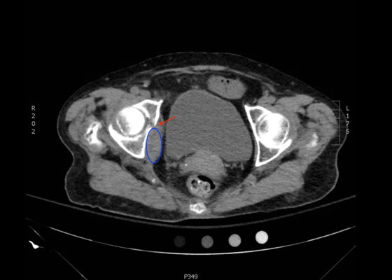
DOI: https://doi.org/10.21980/J8BK8KThe non-contrast CT images show a minimally displaced comminuted fracture of the right acetabulum involving the acetabular roof, medial and anterior walls (red arrows), with associated obturator muscle hematoma (blue oval).
Bedside Ultrasound for the Diagnosis of Small Bowel Obstruction
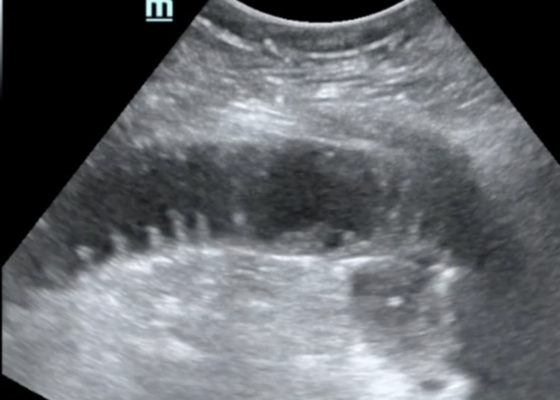
DOI: https://doi.org/10.21980/J86W6PThe POCUS utilizing the low frequency curvilinear probe demonstrates fluid-filled, dilated bowel loops greater than 2.5cm with to-and-fro peristalsis, and thickened bowel walls greater than 3mm, concerning for SBO.
Chancre of Primary Syphilis
DOI: https://doi.org/10.21980/J83342Physical examination revealed a non-tender, erythematous lesion on the glans penis, two similar adjacent satellite lesions, as well as tender inguinal lymphadenopathy. No penile discharge was noted.


