
JETem
  • Home
  • About
    • Aim and Scope
    • Our Team
    • Editorial Board
    • FAQ
  • Issues
    • Current Issue
    • Ahead of Print
    • Past Issues
  • Visual EM
    • Latest Visual EM
    • Search Visual EM
    • Thumbnail Library
  • For Authors
    • Instructions for Authors
    • Submit to JETem
    • Photo Consent
    • Policies
      • Peer Review Policy
      • Copyright Policy
      • Editorial Policy, Ethics and Responsibilities
      • Conflicts of Interest & Informed Consent
      • Open Access Policy
      • Acceptance Fee
  • For Reviewers
    • Instructions for JETem Reviewers
    • Interested in Being a JETem Reviewer?
  • Topic
    • Abdominal/Gastroenterology
    • Administration
    • Board Review
    • Cardiology/Vascular
    • Certifying Exam Cases
      • Clinical Decision-Making
      • Prioritization
      • Communication
      • Procedure & Ultrasound
    • Clinical Informatics, Telehealth and Technology
    • Dermatology
    • EMS
    • Endocrine
    • ENT
    • Faculty Development
    • Genitourinary
    • Geriatrics
    • Hematology/Oncology
    • Infectious Disease
    • Miscellaneous (stats, etc)
    • Neurology
    • Ob/Gyn
    • Ophthalmology
    • Orthopedics
    • Pediatrics
    • Pharmacology
    • Procedures
    • Psychiatry
    • Renal/Electrolytes/Nutrition
    • Respiratory
    • Social Determinants of Health
    • Toxicology
    • Trauma
    • Ultrasound
    • Urology
    • Wellness
    • Wilderness
  • Modality
    • Certifying Exam Practice Cases
      • Clinical Care Cases
      • Communication & Procedural Cases
      • Old School Oral Board Cases
    • Curricula
    • Innovations
    • Lectures
    • Podcasts
    • Simulation
    • Small Group Learning
    • Team Based Learning (TBL)
    • Visual EM
  • Contact Us

Issue 11:3

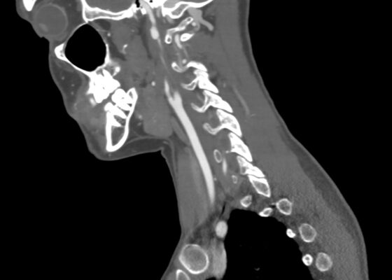
Blunt Cerebrovascular Injury. CT Sagital Internal Carotid Unannotated. JETem 2026

Blunt Cerebrovascular Injury: A Visual Case Report with Computed Tomography Angiography Findings Revealing Carotid and Vertebral Artery Dissection

Edmund Hsu, MD*, Andrew T Kinoshita, MD, MPH^, Elanor Chu, MD† and C Eric McCoy, MD, MPH*

DOI: https://doi.org/10.5070/M5.52229 Issue 11:3[mrp_rating_result]
Computed tomography angiography of the neck showed worsening right ICA dissection with increased size of the pseudoaneurysm to 9 mm and worsening stenosis to greater than 70%. Computed tomography angiography of the neck also showed a new dissection of the V2 segment of the right vertebral artery with a 3 mm pseudoaneurysm and greater than 50% stenosis.
Visual EMCurrent Issue
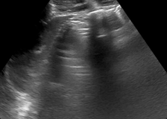
Occult Pneumoperitoneum. Figure 1 Unannotated. JETem 2026

A-Lines below the Diaphragm – A Case Report of Occult Pneumoperitoneum

Yong Hoon Lee MD* and Michael Dalley DO*

DOI: https://doi.org/10.5070/M5.52389Issue 11:3[mrp_rating_result]
The RUQ POCUS focusing over the left liver lobe shows multiple enhanced peritoneal stripe signs (red arrows) and several reverberation artifacts (yellow arrows). The second RUQ POCUS of the right lobe of the liver and the gallbladder shows reverberation artifacts (yellow arrow) and ring-down artifacts (blue arrow). The chest X-ray shows free air under the right diaphragm (orange arrow), which indicates pneumoperitoneum. The CT abdomen and pelvis show pneumoperitoneum near the porta hepatis (green arrow).
Visual EMAbdominal/GastroenterologyCurrent Issue
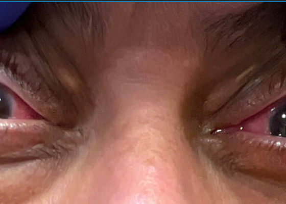
Granulomatosis with Polyangiitis. Chonic Conjunctivitis. JETem 2026

A Case Report of Granulomatosis with Polyangiitis Presenting with Chronic Conjunctivitis

Daniel Martin, PA-C*

DOI: https://doi.org/10.5070/M5.52284Issue 11:3[mrp_rating_result]
Notable bilateral proptosis with conjunctival injection and limbic sparing (clear conjunctiva surrounding the margin of the cornea, shown by the arrow in the image of the eyes) was present.
Visual EMCurrent IssueOphthalmology

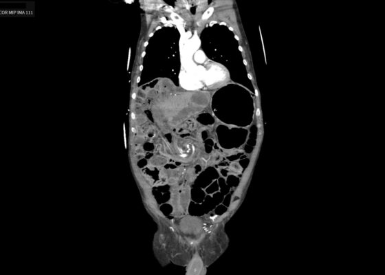
Caught in the Whirl: A Case Report of Midgut Volvulus in an Elderly Patient

Surina Khurana, MD*, Tien Lu, MD^, Carson Dangberg MD^, Kristin Lewis MD, MA^

DOI: https://doi.org/10.5070/M5.52361Issue 11:3[mrp_rating_result]
An upright chest x-ray demonstrated air underneath the diaphragm bilaterally (yellow arrows), enhancing the diaphragmatic contour. A computed tomography angiography (CTA) abdomen and pelvis showed scattered pneumoperitoneum (red arrow), dilated segments of small bowel (orange arrow), and clockwise swirling of the mesentery (blue arrow), concerning for mesenteric volvulus with possible bowel perforation.
Visual EMCurrent Issue
«‹3
Page 3 of 3

JETem is an online, open access, peer-reviewed, journal-repository for EM educators. We are PMC Indexed.

Most Viewed

  • Simple Auricular Hematoma (“Cauliflower Ear”) Task Trainer
  • The Three Breath Method: Teaching Coping Mechanisms for Processing Patient Death
  • Clinical Decision Making: Acute Ischemic Stroke with Large Vessel Occlusion
  • PRIMER: Premedical Introduction to Mentored Emergency Research: Connecting Undergraduate Assistants and Emergency Medicine Residents in a Community Hospital Setting
  • Dangerous Dysrhythmias in the ED: An Interactive EKG Workshop for Emergency Medicine Clerkship Learners

Visit Our Collaborators


Creative Commons Licence
This work is licensed under a Creative Commons Attribution 4.0 International License.

About

Education

Learners should benefit from active learning. JETem accepts submissions of team-based learning, small group learning, simulation, podcasts, lectures, innovations, curricula, question sets, and visualEM.

Scholarship

We believe educators should advance through the scholarship of their educational work. JETem gives educators the opportunity to publish scholarly academic work so that it may be widely distributed, thereby increasing the significance of their results.

Links

  • Home
  • Aim and Scope
  • Current Issue
  • For Reviewers
  • Instructions for Authors
  • Contact Us

Newsletter

Sign up to receive updates from JETem regarding newly published issues and findings.

Copyright Creative Commons Attribution 4.0 International