
Issue 11:3

Car Accident and Respiratory Distress: A Trauma-Based Pneumothorax Simulation Case for Medical Students
DOI: https://doi.org/10.5070/M5.52287By the end of this simulation, learners will be able to: 1) obtain a focused history in the setting of recent motor vehicle accident and respiratory difficulty, 2) identify abnormal respiratory findings consistent with pneumothorax on physical exam, 3) interpret chest x-ray and point-of-care ultrasound to identify the progression of a life-threatening tension pneumothorax,4) assess for potential internal bleeding and hypovolemic shock by integrating imaging findings, vital signs, mental status, and additional components of physical examination, 5) interpret imaging and clinical findings to guide appropriate management decisions, including need for urgent procedural intervention and escalation of care, and 6) deliver a concise oral presentation to an attending physician, including key findings and management plan.
Managing Spontaneous Abortion Hemorrhage: An Adult Simulation Case for Emergency Medicine Residents
DOI: https://doi.org/10.5070/M5.52263By the end of this simulation, learners should be able to: 1) recognize signs of post-abortion hemorrhage, 2) manage hemorrhage using appropriate physical maneuvers, uterotonic medications, and specialist consultation, 3) demonstrate proper placement of a Bakri balloon
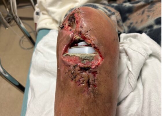

Visual Case Report of a Prosthetic Joint Infection and Chronic Dehiscence following a Total Knee Arthroplasty
DOI: https://doi.org/10.5070/M5.52251Radiographs demonstrated intact prosthetic components (blue) without acute fracture, tibial peri-stem lucency (green) concerning for loosening or infection, as well as joint effusion, and soft-tissue swelling.
Rapidly Progressive Painless Acute Aortic Dissection: A Case Report
DOI: https://doi.org/10.5070/M5.52348A POCUS of the abdomen at that time demonstrated findings concerning for a dissection flap in the abdominal aorta (green arrows), and a new mild to moderate pericardial effusion was seen on a repeat cardiac POCUS suggesting an evolving aortic dissection (blue arrows). The patient’s CTA revealed a type A AAD with extension from the aortic root to the bilateral external and internal iliac vessels as well as the coronary arteries (red arrows).
Computed Tomography Findings of Cerebral Venous Thrombosis: A Case Report
DOI: https://doi.org/10.5070/M5.52331The emergency physician ordered a non-contrast head CT and CT venogram of the head given worsening symptoms with disequilibrium and CVT risk factors of postpartum coagulopathy. The non-contrast head CT demonstrated a hyperdensity occlusion of the left transverse sinus (blue arrow). This serpiginous hyperdensity is called the “cord” or “string” sign, which is thrombosis within the vein. The CT venogram of the head demonstrated occlusion of the left transverse (red arrows) and sigmoid sinus (yellow arrow).
A Case Report of Rare Bilateral Quadriceps Tendon Rupture Diagnosed by Point of Care Ultrasound (POCUS)
DOI: https://doi.org/10.5070/M5.52334Musculoskeletal assessment with POCUS revealed disruption of the left quadriceps tendon (blue arrows) indicated by a large hypoechoic gap (red arrow) with significant surrounding fluid throughout the quadriceps as well as a sizable left knee effusion (orange highlighting). The right lower extremity had a disrupted and retracted quadriceps tendon (yellow arrow) with associated fluid near the injury site and a small right knee joint effusion (orange highlighting). Therefore, he was diagnosed with bilateral quadriceps tendon ruptures.
Hematoma, Hemarthrosis, and Hemotympanum: A Case Report of Acquired Hemophilia A
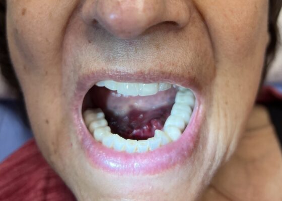
DOI: https://doi.org/10.5070/M5.52344Non-contrast computed tomography (CT) scan of the left knee confirmed a small to moderate suprapatellar joint effusion with Hounsfield unit measurements suggestive of blood products (+47 Hounsfield units), consistent with acute hemarthrosis. The hemarthrosis appears as a gray, fluid-density collection slightly darker than the surrounding tissues that conforms to the suprapatellar space anterior to the femur, continuing distally between the femur and the patella.
A Case Report of a Coin Stack Masquerading as a Button Battery
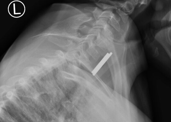
DOI: https://doi.org/10.5070/53886The two images provided are OSH AP and lateral XRs of the patient's neck. The OSH AP view demonstrates a halo sign (green arrow), while the lateral view demonstrates a step-off sign (stick figure). These are key findings with BB ingestions. The halo sign is the presence of two concentric rings on an anterior view XR.