Incidental Hiatal Hernia on Chest X-ray
History of present illness:
A 58-year-old male with history of smoking presented to the emergency department for productive cough, subjective fevers, shortness of breath, and pleuritic chest pain. Patient was discharged three days prior from another hospital where he was admitted for pneumonia and chronic obstructive pulmonary disease (COPD) exacerbation. He was discharged with prescriptions for antibiotics and steroids, which he did not fill. On exam, patient was afebrile and hemodynamically stable. Labs including respiratory syncytial virus and influenza panel were negative.
Significant findings:
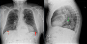
The two-view chest X-ray shows mild opacification of the bilateral lower lobes concerning for pneumonia (red arrows). Incidental retrocardiac opacity with air-fluid level consistent with large hiatal hernia is also observed (green arrow).
Discussion:
A hiatal hernia (HH) is defined as a protrusion of abdominal contents into the thoracic cavity through the diaphragmatic esophageal hiatus.1 HHs can be congenital (1/3000 live births) or, more commonly, acquired.2 HHs are more common in women than in men, and their frequency increases with age, from 10% in patients younger than 40 to 70% in patients over 70 years old.1,3 Acquired HHs can be classified as sliding, paraesophageal, or mixed. Sliding hernias make up greater than 90% of all HHs and occur when the abdominal portion of the esophagus and the cardia & fundus of the stomach slide superiorly through the esophageal hiatus.1 Sliding hernias are clinically significant due to their association with reflux disease.1 Paraesophageal hernias make up less than 10% of all HHs and only involve the stomach fundus passing superiorly into the thoracic cavity.1 Paraesophageal hernias tend to enlarge over time potentially leading to incarceration and subsequent strangulation or perforation.4
Patients with HHs can be asymptomatic or have symptoms such as epigastric fullness, postprandial distress, regurgitation, nausea, chest pain, or cough.4,5 Radiography studies, especially an upper GI barium series, are the preferred examination method to diagnosing HHs with a sensitivity of 77%.6 The primary diagnostic finding is a retrocardiac air-fluid level located within a paraesophageal hernia or intrathoracic stomach.1 Asymptomatic hernias may not require treatment. HH with reflux disease can be managed medically. Symptomatic HHs and paraesophageal hernias might require surgical intervention; thus surgical consultation is recommended.4
In this case, the chest X-ray was concerning for pneumonia, and a large, incidental hiatal hernia was also appreciated. Patient was started on antibiotics for the pneumonia and admitted to internal medicine. The hiatal hernia was not operated on because the patient was asymptomatic.
Topics:
Hiatal hernia, sliding hernia, paraesophageal hernia, retrocardiac air bubble.
References:
- Kahrilas PJ, Kim HC, Pandolfino JE. Approaches to the diagnosis and grading of hiatal hernia. Best Pract Res Clin Gastroenterol. 2008;22(4):601-616. doi: 10.1016/j.bpg.2007.12.007
- Tovar JA. Congenital diaphragmatic hernia. Orphanet J Rare Dis. 2012;7(1):1-15. doi: 10.1186/1750-1172-7-1
- Darby M, Edey A, Chandratreya L, Maskell N. In: Chest X-Ray Interpretation. 1st ed. London, UK: JP Medical Ltd. 2012:163-164.
- Patel S, Shahzad G, Jawairia M, Subramani K, Viswanathan P, Mustacchia P. Hiatus hernia: a rare cause of acute pancreatitis. Case Reports in Medicine. 2016:1-4. doi: 10.1155/2016/2531925
- Pham JT, Spinale RC. Massive incarcerated paraesophageal hiatal hernia. J Am Osteopath Assoc. 2017;117(4):272. doi: 10.7556/jaoa.2017.049
- Weitzendorfer M, Köhler G, Antoniou SA, Pallwein-Prettner L, Manzenreiter L, Schredl P, et al. Preoperative diagnosis of hiatal hernia: barium swallow X-ray, high-resolution manometry, or endoscopy? Eur Surg. 2017;49(5):210-217. doi:10.1007/s10353-017-0492-y


