
JETem
  • Home
  • About
    • Aim and Scope
    • Our Team
    • Editorial Board
    • FAQ
  • Issues
    • Current Issue
    • Ahead of Print
    • Past Issues
  • Visual EM
    • Latest Visual EM
    • Search Visual EM
    • Thumbnail Library
  • For Authors
    • Instructions for Authors
    • Submit to JETem
    • Photo Consent
    • Policies
      • Peer Review Policy
      • Copyright Policy
      • Editorial Policy, Ethics and Responsibilities
      • Conflicts of Interest & Informed Consent
      • Open Access Policy
      • Acceptance Fee
  • For Reviewers
    • Instructions for JETem Reviewers
    • Interested in Being a JETem Reviewer?
  • Topic
    • Abdominal/Gastroenterology
    • Administration
    • Board Review
    • Cardiology/Vascular
    • Certifying Exam Cases
      • Clinical Decision-Making
      • Prioritization
      • Communication
      • Procedure & Ultrasound
    • Clinical Informatics, Telehealth and Technology
    • Dermatology
    • EMS
    • Endocrine
    • ENT
    • Faculty Development
    • Genitourinary
    • Geriatrics
    • Hematology/Oncology
    • Infectious Disease
    • Miscellaneous (stats, etc)
    • Neurology
    • Ob/Gyn
    • Ophthalmology
    • Orthopedics
    • Pediatrics
    • Pharmacology
    • Procedures
    • Psychiatry
    • Renal/Electrolytes/Nutrition
    • Respiratory
    • Social Determinants of Health
    • Toxicology
    • Trauma
    • Ultrasound
    • Urology
    • Wellness
    • Wilderness
  • Modality
    • Certifying Exam Practice Cases
      • Clinical Care Cases
      • Communication & Procedural Cases
      • Old School Oral Board Cases
    • Curricula
    • Innovations
    • Lectures
    • Podcasts
    • Simulation
    • Small Group Learning
    • Team Based Learning (TBL)
    • Visual EM
  • Contact Us

Visual EM

Creative Commons images

An Unusual Case of Hematemesis

Amanda Amen, BA*, Jane Xiao, MD^, Julie Parks-Bortel, MD† and Shanna Jones, MD†

DOI: https://doi.org/10.21980/J84H00Issue 3:4[mrp_rating_result]
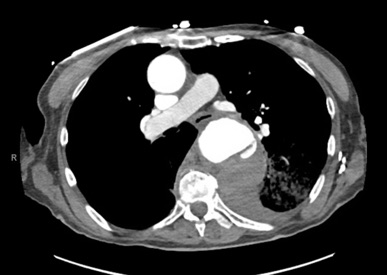
The patient’schest X-ray revealed a prominent mediastinum and opacification in the left middle and lower lung fields. The CT showed an aortic aneurysm extending from the thorax to the abdomen with rupture near T7 (blue arrow). It also showed periaortic hemorrhage with active extravasation (green arrow) likely secondary to a penetrating ulcer and bilateral pulmonary opacities concerning for hemothorax (pink arrow).
Cardiology/VascularVisual EM
Creative Commons images

Extensive Aortic Dissection with Normal Vital Signs

Meryl Abrams, MD*, Nicole Pagliuso, EMT* and Xiao Chi Zhang, MD, MS*

DOI: https://doi.org/10.21980/J80S6SIssue 3:4[mrp_rating_result]
The patient was found to have a Stanford type A dissection (see yellow arrow) with visible false lumen starting at aortic arch (see green circle). The dissection extended into the descending aorta (see blue circle) as shown by the false lumen (red highlighted area) visible on CT. The radiologist performed a reconstruction of the aorta, which showed that the left kidney was not being perfused, making the kidney not visible on the reconstruction.
Cardiology/VascularVisual EM
Creative Commons images

Croup

Victor Cisneros, MD, MPH*, Joseph Zakaria, BS* and Wirachin Hoonpongsimanont, MD*

DOI: https://doi.org/10.21980/J8W05JIssue 3:4[mrp_rating_result]
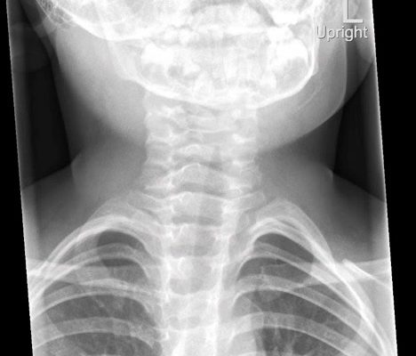
The anteroposterior X-ray reveals the classic steeple sign (blue outline) indicative of subglottic edema leading to tracheal narrowing, consistent with croup. The lateral x-ray shows narrowing of the subglottic region (green outline and arrows).

PediatricsRespiratoryVisual EM
Creative Commons images

Various Degrees of Thermal Burns

Hamid Ehsani-Nia, DO*, Mohammad I Ehsani-Nia, BS* and John Collins, MD*

DOI: https://doi.org/10.21980/J8R91WIssue 3:4[mrp_rating_result]
On exam,there is a large swath of skin with evidence of thermal injury involving the neck, shoulder, chest, and face, including damage to the ear, external nostril, and lips. Burns exhibit varying degrees of severity and total approximately 4.5% of the body surface area. Several areas are charred and insensate to pinprick. The left earlobe is partially burned off. Patient's airway is patent with no evidence of thermal injury or obstruction to the oropharynx or nasal vestibule.
TraumaVisual EM
Creative Commons images

Pemphigoid Gestationis

Michael Niechayev, BS* and Lauren Sylwanowicz, MD*

DOI: https://doi.org/10.21980/J8MG9DIssue 3:4[mrp_rating_result]
Physical exam findings were significant for 1-3 cm diameter well-demarcated superficial ulcers on the patient’s abdomen and extremities, with mucosal sparing. Several small tense bullae were present on the bilateral inner thighs and numerous small reddish plaques were scattered over the patient’s back. Nikolsky’s sign was negative. No lymphadenopathy was noted.
DermatologyVisual EM
Creative Commons images

Don’t Forget the Pacemaker – A Rare Complication

Amanda Esposito, MD*, Kenneth Rapp, MD* and Albert Ritter, MD*

DOI: https://doi.org/10.21980/J8GS7HIssue 3:4[mrp_rating_result]
The ECG demonstrated the presence of pacemaker spikes without appropriate capture (green arrows) and a ventricular escape rhythm which can be identified by an absence of P waves prior to the QRS complex (purple arrows). The portable chest X- demonstrated displaced pacemaker leads (red arrows) that were coiled around the pulse generator (blue arrow).   
Cardiology/VascularVisual EM
Creative Commons images

Bedside Ultrasound of Retinal Detachment in a 19-year-old

Rami San Gabriel, BS*, Maili Alvarado, MD* and Vy Han, MD*

DOI: https://doi.org/10.21980/J80W6TIssue 3:3[mrp_rating_result]
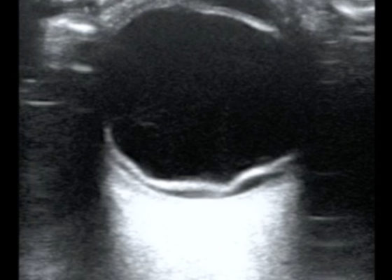
The ocular point of care ultrasound (POCUS) utilizing a high frequency linear probe shows a retinal detachment (RD) with a thick, hyperechoic undulating membrane in the vitreous humor that is anchored at the ora serrata anteriorly and the optic disc posteriorly. Note that the retina is detached all the way to the optic disc making it "mac off." The macula, and more specifically the fovea, is located in the central retina and contains a high concentration of cone photoreceptors responsible for central, high resolution, color vision. In a "mac on" RD, the retina detaches in the periphery but remains intact centrally. This is an ophthalmologic emergency and timely diagnosis and intervention can be vision saving. This patient also has evidence of a posterior vitreous hemorrhage which has a characteristic swirling appearance with kinetic exam on real-time imaging. The detached vitreous body is not as well defined and is not anchored posteriorly to the optic disc.
OphthalmologyVisual EM
Creative Commons images

Bullous Emphysema

Valerie Lew, MD*, John Costumbrado, MD, MPH^ and Steven Kim, MD^

DOI: https://doi.org/10.21980/J8W62GIssue 3:3[mrp_rating_result]
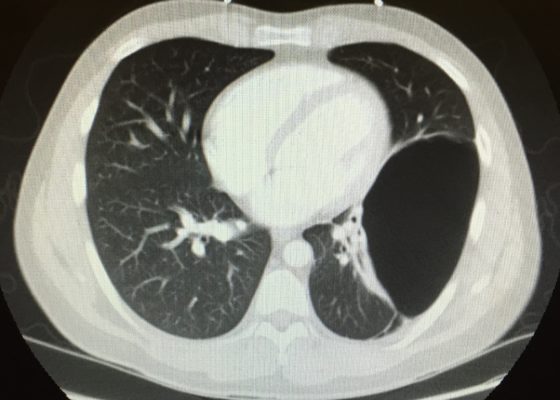
The upright chest X-ray shows a large lucent area in the left lower lung field without lung markings, with associated curvilinear opacities (yellow arrows) consistent with a large air-filled bulla. The bulla is large enough to compress adjacent lung tissue as shown by the visible pleural line (blue line). The discontinuity of the pleural line and presence of lung markings superiorly makes these findings more consistent with bulla than pneumothorax. The chest computed tomography (CT) confirmed a large left hemithorax bulla.
RespiratoryVisual EM
«‹28›»
Page 28 of 45

JETem is an online, open access, peer-reviewed, journal-repository for EM educators. We are PMC Indexed.

Most Viewed

  • Simple Auricular Hematoma (“Cauliflower Ear”) Task Trainer
  • The Three Breath Method: Teaching Coping Mechanisms for Processing Patient Death
  • Clinical Decision Making: Acute Ischemic Stroke with Large Vessel Occlusion
  • PRIMER: Premedical Introduction to Mentored Emergency Research: Connecting Undergraduate Assistants and Emergency Medicine Residents in a Community Hospital Setting
  • Dangerous Dysrhythmias in the ED: An Interactive EKG Workshop for Emergency Medicine Clerkship Learners

Visit Our Collaborators


Creative Commons Licence
This work is licensed under a Creative Commons Attribution 4.0 International License.

About

Education

Learners should benefit from active learning. JETem accepts submissions of team-based learning, small group learning, simulation, podcasts, lectures, innovations, curricula, question sets, and visualEM.

Scholarship

We believe educators should advance through the scholarship of their educational work. JETem gives educators the opportunity to publish scholarly academic work so that it may be widely distributed, thereby increasing the significance of their results.

Links

  • Home
  • Aim and Scope
  • Current Issue
  • For Reviewers
  • Instructions for Authors
  • Contact Us

Newsletter

Sign up to receive updates from JETem regarding newly published issues and findings.

Copyright Creative Commons Attribution 4.0 International