
Respiratory
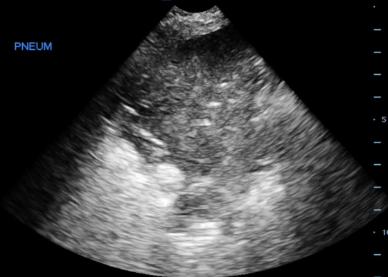

Pneumonia Diagnosed by Point-of-Care Ultrasound
DOI: https://doi.org/10.21980/J8XP64Point-of-care ultrasound of the left lower lobe demonstrates lung hepatization, a classic finding for pneumonia. In addition, a shred sign is present with both air bronchograms and focal B-lines—all suggestive of poorly aerated, consolidated lung. The patient was started on antibiotics and admitted to the hospital with a diagnosis of community-acquired pneumonia.

Re-expansion Pulmonary Edema
DOI: https://doi.org/10.21980/J8WS6VInitial chest X-ray (chest X-ray 1) showed a right-sided pleural effusion with compressive atelectasis of the mid to lower right lung. Repeat chest X-ray immediately after evacuation (chest X-ray 2) shows improvement of the pleural effusion and a new trace apical right pneumothorax measuring 6.7 mm. When the patient became tachypneic, a third X-ray (chest X-ray 3) showed persistent trace apical right pneumothorax measuring 6.7 mm.

Open Pneumothorax
DOI: https://doi.org/10.21980/J88036A large chest wound was clinically obvious. A chest radiograph performed after intubation showed subcutaneous emphysema, an anterior rib fracture, and a right-sided pneumothorax. He was then taken to the operating room for further management.
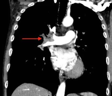
Acute, massive pulmonary embolism with right heart strain and hypoxia requiring emergent tissue plasminogen activator (TPA) infusion
DOI: https://doi.org/10.21980/J84K5KCT angiogram showed multiple large acute pulmonary emboli, most significantly in the distal right main pulmonary artery (image 1 and 2). Additional pulmonary emboli were noted in the bilateral lobar, segmental, and subsegmental levels of all lobes. There was a peripheral, wedge-shaped consolidation surrounded by groundglass changes in the posterolateral basal right lower lobe that was consistent with a small lung infarction (image 3).

Ventilator TBL
DOI: https://doi.org/10.21980/J8H01MAt completion of this TBL the learner should be able to: 1. Choose appropriate ventilator settings and adjustments for a patient with lung injury or obstructive lung pattern 2. Troubleshoot an alarming ventilator 3. Set up the most common ventilator in their ED 4. Set up the equipment needed for transtracheal jet ventilation.

An Elderly Female with Dyspnea and Abdominal Pain
DOI: https://doi.org/10.21980/J83S3KRadiography shows a dilated, gas-filled structure that fills nearly the entire left hemi-thorax. Lung markings are visible in the uppermost portion of the left hemi-thorax. There is mediastinal shift to the right. In the visualized portion of the abdomen, dilated loops of bowel are also visualized. This constellation of findings is consistent with a tension gastrothorax.
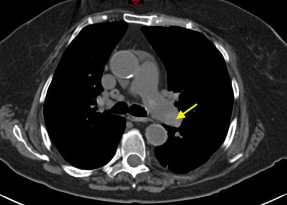
Pulmonary Embolism: Diagnosis by Computerized Tomography without Intravenous Contrast
DOI: https://doi.org/10.21980/J8001ZNon-contrast CT of the chest demonstrates hyper-densities within both central and sub-segmental pulmonary arteries bilaterally (see yellow arrows). The right ventricle is dilated.

Large Right Pleural Effusion
DOI: https://doi.org/10.21980/J8D59FChest x-ray and bedside ultrasound revealed a large right pleural effusion, estimated to be greater than two and a half liters in size.