
Neurology

Novel Emergency Medicine Curriculum Utilizing Self-Directed Learning and the Flipped Classroom Method: Neurologic Emergencies Small Group Module
DOI: https://doi.org/10.21980/J89H0JWe aim to teach the presentation and management of cardiovascular emergencies through the creation of a flipped classroom design. This unique, innovative curriculum utilizes resources chosen by education faculty and resident learners, study questions, real-life experiences, and small group discussions in place of traditional lectures. In doing so, a goal of the curriculum is to encourage self-directed learning, improve understanding and knowledge retention, and improve the educational experience of our residents.

Spontaneous Intracranial Hemorrhage in Severe Hemophilia A: A Rare Cause of Seizure in a Young Child
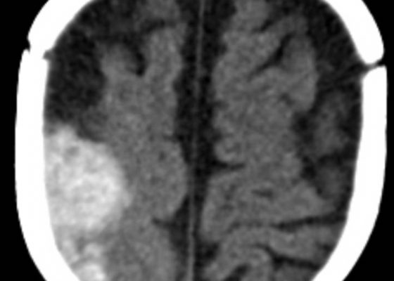
DOI: https://doi.org/10.21980/J8G91DA computed tomography (CT) scan of the head without contrast was obtained out of concern for intracranial pathology due to the patient’s young age and the witnessed focal seizure. The CT showed a 4.2 x 1.2 x 1.5 cm acute extra-axial intracranial right frontoparietal hemorrhage favoring epidural over subdural hemorrhage given its lenticular shape. There was no underlying fracture, herniation or midline shift identified.

Acute Ischemic Stroke
DOI: https://doi.org/10.21980/J8R04XBy the end of this simulation session, learners will be able to: 1) recognize a CVA using the National Institutes of Health Stroke Scale (NIHSS), 2) understand and properly utilize the NIHSS, 3) list appropriate imaging and laboratory orders for a CVA work-up, 4) determine appropriate subspecialty consultation, 5) discuss common stroke syndromes and associated cerebral locations, 6) review indications and contraindications for tissue plasminogen activator (tPA), 7) review hospital specific stroke protocol.

A Woman with Arm Spasms
DOI: https://doi.org/10.21980/J8VP88The patient had a witnessed episode of isolated left upper extremity jerking, shown in the video, during which she was completely awake and conversant. Lab results were significant for serum glucose of 1167 mg/dL, no anion gap, and negative serum/urine ketones. She had a computed tomography (CT) of the head that did not show any acute pathology, and underwent a brain magnetic resonance imaging (MRI) without any signs of stroke or other pathology, shown below.
Beware the Devastating Outcome of a Common Procedure
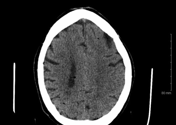
DOI: https://doi.org/10.21980/J8T336Non-contrast head computed tomography (CT) demonstrates multifocal bilateral hypodense lesions (white arrows) representing air emboli. Note the lesions are located in the intra-axial distribution which indicates an underlying vascular origin.
Guillain-Barrè
DOI: https://doi.org/10.21980/J8TH06At the conclusion of the simulation session, learners will be able to: 1) Recognize the clinical signs and symptoms associated with Guillain-Barré syndrome, including muscle weakness and hyporeflexia. 2) Identify abnormal vital signs secondary to dysautonomia. 3) Discuss evaluation for impending respiratory failure, including bedside pulmonary function testing. 4) Discuss the management of Guillain-Barré, including management of dysautonomia and respiratory failure, as well as definitive management with plasmapheresis versus intravenous immunoglobulin. 5) Appropriately disposition the patient to the intensive care unit. 6) Effectively communicate with team members and nursing staff during resuscitation of a critically ill patient
Radial Nerve Palsy
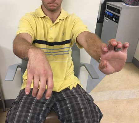
DOI: https://doi.org/10.21980/J8KS7FOn physical exam, the patient was unable to extend his right wrist, thumb, and fingers, and had no sensation of his 1stdorsal interosseous muscles up to the proximal dorsal radial aspect of his forearm. The patient also had slight weakness in thumb abduction. Triceps strength was preserved.