
Posts by JETem

An Unusual Case of Hematemesis
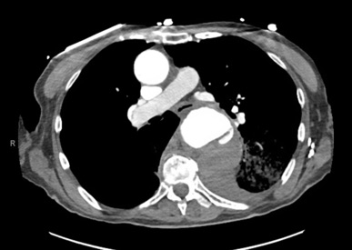
DOI: https://doi.org/10.21980/J84H00The patient’schest X-ray revealed a prominent mediastinum and opacification in the left middle and lower lung fields. The CT showed an aortic aneurysm extending from the thorax to the abdomen with rupture near T7 (blue arrow). It also showed periaortic hemorrhage with active extravasation (green arrow) likely secondary to a penetrating ulcer and bilateral pulmonary opacities concerning for hemothorax (pink arrow).

Extensive Aortic Dissection with Normal Vital Signs
DOI: https://doi.org/10.21980/J80S6SThe patient was found to have a Stanford type A dissection (see yellow arrow) with visible false lumen starting at aortic arch (see green circle). The dissection extended into the descending aorta (see blue circle) as shown by the false lumen (red highlighted area) visible on CT. The radiologist performed a reconstruction of the aorta, which showed that the left kidney was not being perfused, making the kidney not visible on the reconstruction.
Croup
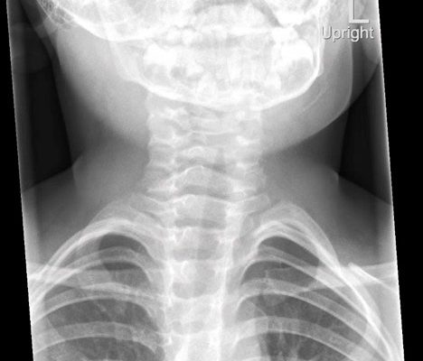
DOI: https://doi.org/10.21980/J8W05JThe anteroposterior X-ray reveals the classic steeple sign (blue outline) indicative of subglottic edema leading to tracheal narrowing, consistent with croup. The lateral x-ray shows narrowing of the subglottic region (green outline and arrows).
Various Degrees of Thermal Burns
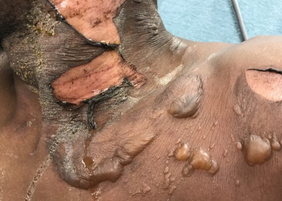
DOI: https://doi.org/10.21980/J8R91WOn exam,there is a large swath of skin with evidence of thermal injury involving the neck, shoulder, chest, and face, including damage to the ear, external nostril, and lips. Burns exhibit varying degrees of severity and total approximately 4.5% of the body surface area. Several areas are charred and insensate to pinprick. The left earlobe is partially burned off. Patient's airway is patent with no evidence of thermal injury or obstruction to the oropharynx or nasal vestibule.

Pemphigoid Gestationis
DOI: https://doi.org/10.21980/J8MG9DPhysical exam findings were significant for 1-3 cm diameter well-demarcated superficial ulcers on the patient’s abdomen and extremities, with mucosal sparing. Several small tense bullae were present on the bilateral inner thighs and numerous small reddish plaques were scattered over the patient’s back. Nikolsky’s sign was negative. No lymphadenopathy was noted.

Don’t Forget the Pacemaker – A Rare Complication
DOI: https://doi.org/10.21980/J8GS7HThe ECG demonstrated the presence of pacemaker spikes without appropriate capture (green arrows) and a ventricular escape rhythm which can be identified by an absence of P waves prior to the QRS complex (purple arrows). The portable chest X- demonstrated displaced pacemaker leads (red arrows) that were coiled around the pulse generator (blue arrow).

Novel Emergency Medicine Curriculum Utilizing Self-Directed Learning and the Flipped Classroom Method: Cardiovascular Emergencies Small Group Module
DOI: https://doi.org/10.21980/J8X334We aim to teach the presentation and management of cardiovascular emergencies through the creation of a flipped classroom design. This unique, innovative curriculum utilizes resources chosen by education faculty and resident learners, study questions, real-life experiences, and small group discussions in place of traditional lectures. In doing so, a goal of the curriculum is to encourage self-directed learning, improve understanding and knowledge retention, and improve the educational experience of our residents.

The Continuous Residency Improvement Committee (CRIC) – A Novel Twist for Program Evaluation in an Academic Emergency Medicine Residency Program
DOI: https://doi.org/10.21980/J8SD17The purpose of this innovation was to develop a novel approach to continuous program evaluation and improvement using a multisource feedback design to improve resident satisfaction with the program’s responsiveness to feedback while addressing the ACGME mandate for self-study.