
Posts by JETem
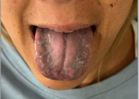

Woman with a Blackened Tongue: A Case Report
DOI: https://doi.org/10.5070/M5.52329The patient’s vital signs were blood pressure 109/72 mmHg, respiratory rate 18 breaths per minute, pulse 93 beats per minute, temperature 97°F. Physical examination revealed notable black discoloration of the tongue and gingiva, along with hyperpigmented skin exhibiting numerous diffuse nevi distributed across the face and upper extremities.
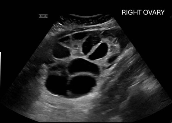
A Woman’s Infertility Journey Complicated by Severe Ovarian Hyperstimulation Syndrome – A Case Report
DOI: https://doi.org/10.5070/M5.52309Computed tomography (CT) of the abdomen and pelvis demonstrated pleural effusions in the lung bases (blue stars), ascites (blue stars), and enlarged ovaries with multiple cysts/follicles (white arrows). A formal pelvic US also demonstrated large volume ascites (white diamonds) and bilateral ovarian enlargement with numerous cysts. A CT angiogram (CTA) of the chest demonstrated small bilateral pleural effusions and no obvious pulmonary embolism.
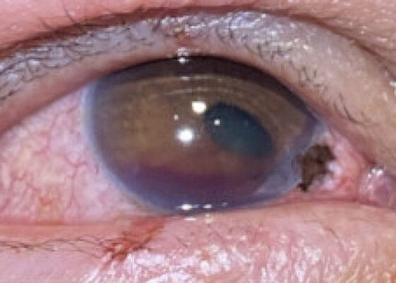
A Case Report of Corneoscleral Laceration with Open Globe Injury and Iris Prolapse
DOI: https://doi.org/10.5070/M5.52326Closer examination of the right eye revealed a 2 mm x 1 mm corneoscleral laceration to the 4-5 o'clock position at the border of zone I (yellow highlighting) and II (blue highlighting) with iris prolapse (yellow star) and plugging. Additionally, the affected eye revealed a peaked pupil (cyan highlighting) in the inferonasal direction with associated corectopia and a 2 mm Grade I hyphema (pink highlighting). Immediately inferior to the corneal laceration, 1 mm epithelial injury of the lower palpebra was present (green cross). Fluorescein exam demonstrated uptake at the 4-5 o'clock position (yellow arrow) but was negative for Seidel’s sign.
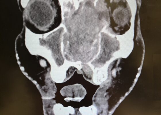
Diagnosis of Sinonasal Carcinoma in the Emergency Department: A Case Report Highlighting Red Flag Symptoms
DOI: https://doi.org/10.5070/M5.52257Physical exam revealed a gross deformity of the left side of the face with soft tissue swelling with no overlying skin changes. The left eye was proptotic and completely immobile, with cranial nerve (CN) 3, 4, and 6 palsies. Additionally, a large obstructive mass was noted in the left naris with resultant rightward displacement of the nasal septum. Intraocular pressures (IOPs) were measured to be 6 mmHg in the right eye and 11 mmHg in the left eye.
Imaging, including maxillofacial and neck soft tissue CTs revealed a large, destructive, soft tissue mass centered in the nasal cavity with significant osseous destruction of the midface and skull base (red highlighted area). There was mild intracranial extension with the mass abutting or infiltrating the inferior frontal lobes (green highlighted area). The nasal cavity and nasopharynx were obstructed (orange highlighted area). The mass invaded the left orbit with associated left-sided proptosis, left globe deformity, and compressed the left optic nerve and the left optic chiasm (blue arrows).
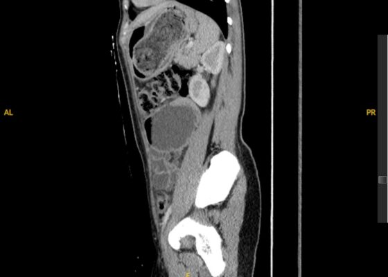
Trapped In Transit – A Case Report of a Pediatric Gastric Bezoar Causing Intermittent Small Bowel Obstruction
DOI: https://doi.org/10.5070/M5.52259The CT scan identified a heterogenous, mass-like lesion extending from the gastric fundus to the antrum, along with a small bowel obstruction (SBO) and significant small bowel dilation. An additional smaller heterogenous mass-like lesion was seen in the small bowel, causing the SBO. Given the mass-like lesion’s mixed attenuation and presence of internal air, a gastric bezoar was strongly suspected.
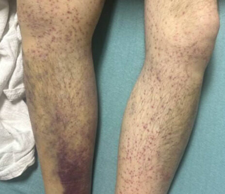
Overlooked and Undernourished: A Case Report of Scurvy Linked to Food Insecurity
DOI: https://doi.org/10.5070/M5.52313Images A and B of the lower extremities show what initially appeared to be a petechial rash with scattered ecchymoses bilaterally, primarily on the right lower leg. On closer examination, images C and D more clearly show these spots to be flat and non-blanching, indicative of follicular hyperkeratosis with perifollicular hemorrhages. The classic coiled or corkscrew hair findings are not seen in these images. Image E shows evidence of palatal ecchymosis, without evidence of gingival bleeding, likely due to loss of dentition. This constellation of exam findings reflects the production of weakened or dysfunctional collagen in connective tissue and vascular structures and is strongly suggestive of the clinical diagnosis of scurvy. Image F shows resolution of the patient’s dermatologic findings after three weeks of treatment.

Enhancing Emergency Medicine Resident Education: A Weekly Education Series to Augment Electrocardiogram Education
DOI: https://doi.org/10.5070/M5.52141The goals of the curriculum were to establish an asynchronous ECG curriculum to help improve standardization of EM resident education, expose EM residents to a more comprehensive ECG curriculum, increase active learning in this arena, and decrease the administrative burden while not dedicating further conference time towards ECG education.

Development and Design of a Pediatric Case-Based Virtual Escape Room on Acute Iron Toxicity
DOI: https://doi.org/10.5070/M5.52192By the end of the activity, learners should be able to: 1)recognize the history and clinical presentation of acute iron toxicity; 2) demonstrate knowledge of the necessary workup in suspected iron toxicity; 3) identify the stages of acute iron toxicity; 4) identify management of iron toxicity and its complications; 5) perform appropriate management in the setting of decompensated hemorrhagic shock and hypovolemia; and 6) demonstrate teamwork through communication and collaboration.