
Posts by JETem


Unravelling the Mystery of a Continuous Coil: A Case Report
DOI: https://doi.org/10.21980/J8PM00A CT scan of the abdomen and pelvis with intravenous contrast for evaluation of new onset abdominal pain and distension was obtained in the emergency department. The axial view (CT Image A) shows the coil pack from the prior coil-assisted retrograde transvenous obliteration procedure, seen in the left renal vein and gastric varix (red arrow). The path of the coil (yellow arrow) is continuous into the inferior vena cava (CT Image B). It is then seen (CT Image C) situated in the right ventricle (green arrow). Finally, the coil pack is seen in a coronal section, demonstrating its upward path (blue arrow) in the inferior vena cava. (CT Image D). Additional findings included ascites with advanced cirrhosis. As noted in the CT images, a vascular embolization coil was seen within a varix near the junction of the left renal vein. This appeared to have unraveled and extended superiorly into the inferior vena cava and ultimately into the right atrium and right ventricle.

Clinical and Radiologic Features of Fulminant Pediatric Autoimmune Encephalitis: A Case Report
DOI: https://doi.org/10.21980/J8JW75The neurology service was consulted in the ED and multisequence MRI and MR angiography (MRA) of the brain were obtained without and with IV contrast. Diffusion-weighted imaging (DWI) and T2-weighted-Fluid-Attentuated Inversion Recovery (FLAIR) sequences showed multifocal small areas of diffusion signal abnormality in the brainstem and basal ganglia (red asterisks) suggestive of ischemia. Additional multifocal bilateral supra- and infratentorial foci of signal abnormality including subcortical white matter and deep grey matter were highly concerning for encephalitis or demyelinating disease. MRI was repeated on day 3 and day 7 during evolution of disease.

Case Report of a Pelvic Kidney with Ureteral Obstruction from Inguinal Hernia Entrapment and Concurrent Cryptorchid Testis
DOI: https://doi.org/10.21980/J8F345The patient was afebrile with normal lactate and white blood cell count. Initial CT imaging showed an ectopic right pelvic kidney with entrapment of his right ureter within an indirect right inguinal hernia causing severe hydronephrosis (coronal: white arrow). Also discovered was an ovoid hypodensity in the right anterior pelvis consistent with right undescended testis (axial: orange arrow; coronal: green arrow) that was previously unknown to the patient, with a normal left scrotal testis (axial: red arrowhead; coronal: blue arrowhead). Other potential etiologies of the patient’s symptoms could include appendicitis or incarcerated inguinal hernia, though the imaging results and absence of systemic inflammatory response syndrome made these causes less likely.

Syncope Due to a Ruptured Ectopic Pregnancy
DOI: https://doi.org/10.21980/J86M0NAt the conclusion of this simulation, the learner will be able to: 1) review the initial management of syncope; 2) utilize laboratory and imaging techniques to diagnose a ruptured ectopic pregnancy; and 3) demonstrate the ability to resuscitate and disposition an unstable ruptured ectopic pregnancy.

Massive Upper Gastrointestinal Bleeding
DOI: https://doi.org/10.21980/J8W93WBy the end of this simulation, learners will be able to: 1) manage a hypotensive patient with syncope and hematemesis, 2) pharmacologically manage an acute UGIB addressing the various causes, 3) recognize worsening clinical status and intervene by performing difficult airway management, 4) place a gastroesophageal balloon tamponade device.
Principles of Hypotensive Shock: A Video Introduction to Pathophysiology and Treatment Strategies
DOI: https://doi.org/10.21980/J8MS84By the end of this module, participants should be able to: 1) review basic principles of cardiovascular physiology; 2) describe the 4 general pathophysiologic mechanisms of hypotensive shock; 3) recognize various etiologies for each mechanism of hypotensive shock; 4) recognize differences in the clinical presentation of each mechanism of hypotensive shock; 5) cite the basic approach to treatment for each mechanism of hypotensive shock.
A Lecture to Teach an Approach and Improve Resident Comfort in Leading Resuscitation of Young Infants in the Emergency Department
DOI: https://doi.org/10.21980/J8H36JBy the end of this lecture, participants should be able to: 1) apply a consistent approach to the initial resuscitation of a critically ill young infant in the emergency department; 2) select appropriate medications and equipment for use in resuscitation of critically ill young infants; 3) describe the components of the Pediatric Assessment Triangle,6 which can be used to identify critically ill infants and children; 4) improve comfort in resuscitating young infants in the emergency department.
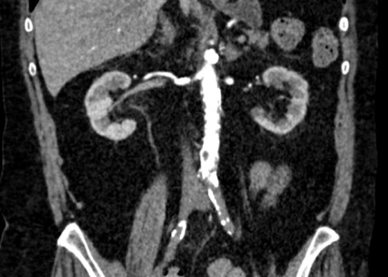
Initial Management and Recognition of Aortoiliac Occlusive Disease, A Case Report
DOI: https://doi.org/10.21980/J87M0ZComputerized tomography with angiography (CTA) of the entire aorta demonstrated an occluded distal infrarenal aorta with extension into the bilateral common femoral arteries (red outline), lack of flow through femoral arteries (yellow outline) and trickle flow reconstituted distally consistent with aortoiliac occlusive disease (blue outline). Some small segments of the proximal celiac axis showed signs of occlusion (purple outline). A short segment of non-specific bowel wall thickening, which may have been related to ischemic changes, was also seen (not seen on images). The included coronal slice shows the extent of the bilateral occlusive burden, with three-dimensional reconstruction emphasizing the same findings.