Takotsubo (Stress) Cardiomyopathy
History of present illness:
A 59-year-old male presented to the emergency department in shock from pneumonia. The patient was initially afebrile, pulse rate 120 beats per minute, blood pressure 117/69 mmHg, respiratory rate 42 breaths per minute, pulse oximetry 94% on a non-rebreather mask and a lactate of 14 mmol/L. He became progressively more hypotensive despite fluid resuscitation and was started on norepinephrine. Shortly after, the patient developed torsades de pointes that was terminated with intravenous push magnesium. His initial ECG had shown sinus tachycardia; however, repeat ECG showed ST-segment elevation in the inferolateral leads and the patient had troponin I elevation that peaked at 16 ng/mL.
Significant findings:
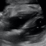
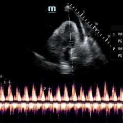
Bedside echocardiography showed the findings consistent with Takotsubo cardiomyopathy. Echocardiographic images are shown in subxiphoid (A) and apical four chamber (B) views. Note the apical ballooning appearance (asterisk) of the left ventricle (LV).
Discussion:
Formal echocardiography confirmed features classic for Takotsubo (stress) cardiomyopathy, including globally depressed left ventricle (LV) ejection fraction, systolic apical ballooning appearance of the LV, mid and apical segments of LV depression, and hyper kinesis of the basal walls. Takotsubo cardiomyopathy is a syndrome known to cause ST-segment elevation on ECG, transient LV dysfunction, and dysrhythmia in the absence of acute obstructive coronary disease. There is no consensus on diagnostic criteria; however, these criteria are commonly used: 1) transient hypokinesis, akinesis, or dyskinesis in the LV mid-segments with or without apical involvement; regional wall motion abnormalities that extend beyond a single epicardial vascular distribution; and frequently, but not always, a stressful trigger 2) the absence of acute coronary disease or angiographic evidence of acute plaque rupture 3) new ECG abnormalities (ST-segment elevation and/or T-wave inversion) or modest elevation in cardiac troponin, and 4) the absence of myocarditis or pheochromocytoma.1 This syndrome is more common in women, older adults, and in patients presenting with neurologic pathology.2 It is crucial to appreciate how common transient cardiomyopathy is during critical illness and sepsis, and the importance of screening for cardiac dysfunction. It has been well described in the literature that up to one-third of patients in septic shock will suffer from transient global cardiomyopathy, thought to be caused by a number of mechanisms including inflammatory mediators, catecholamine excess, microvascular dysfunction and perturbations of the brain-heart axis.2-5 Management is supportive, using a standard approach to managing patients with heart failure and cardiomyopathy. The majority of patients will recover systolic function within several days to weeks.4
Topics:
Bedside ultrasound, echocardiography, septic shock, cardiomyopathy, cardiology.
References:
- Akashi YJ, Goldstein DS, Barbaro G, Ueyama T. Takotsubo cardiomyopathy: a new form of acute, reversible heart failure. Circulation. 2008;118(25):2754-2762. doi: 10.1161/CIRCULATIONAHA.108.767012
- Templin C, Ghadri JR, Diekmann J, Napp LC, Bataiosu DR, Jaguszewski M, et al. Clinical features and outcomes of Takotsubo (stress) cardiomyopathy. N Engl J Med. 2015;373(10):929-938. doi: 10.1056/NEJMoa1406761
- Wittstein IS, Thiemann DR, Lima JA, Baughman KL, Schulman SP, Gerstenblith G, et al. Neurohumoral features of myocardial stunning due to sudden emotional stress. N Engl J Med. 2005;352(6):539-548. doi: 10.1056/NEJMoa040346
- Gianni M, Dentali F, Grandi AM, Sumner G, Hiralal R, Lonn E.Apical ballooning syndrome or takotsubo cardiomyopathy: a systematic review. Eur Heart J. 2006;27(13):1523-1529. doi: 10.1093/eurheartj/ehl032
- Nef HM, Mollmann H, Kostin S, Troidl C, Voss S, Weber M, et al. Tako-Tsubo cardiomyopathy: intraindividual structural analysis in the acute phase and after functional recovery. Eur Heart J. 2007;28(20):2456-2464. doi: 10.1093/eurheartj/ehl570


