
Ultrasound


Ultrasonographic Findings of Acute Achilles Tendon Rupture
DOI: https://doi.org/10.21980/J8063SThe ultrasound video clip shows a longitudinal view of the AT during a dynamic exam. While the patient’s foot is gently passively dorsi/plantar flexed, the video demonstrates first a normal Achilles tendon (from the unaffected extremity) without disruption (highlighted by a yellow bracket on screen left). Then it shows an abnormal tendon (the patient’s affected side) with disruption of the typical linear tendon fibers (red arrow identifies area of rupture). Dynamic testing shows the movement of the distal stump of the AT is evident adjacent to hypoechoic fluid that is reactive edema or blood from the acute rupture.
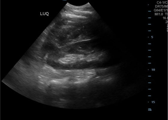
FAST Exam to Diagnose Subcapsular Renal Hematoma
DOI: https://doi.org/10.21980/J8NP8DA bedside point of care ultrasound FAST exam was performed revealing a left subcapsular renal hematoma. The hematoma was a non-compressing hematoma, evidenced by preserved renal contour with the hematoma labeled with a red H and the normal renal contour labeled with a green K. Additionally, cortical necrosis and ischemia can be characterized by a dark, hypoechogenic renal cortex on ultrasonography with a decrease in flow to the cortex on color doppler which was not seen on this patient, providing further evidence that the hematoma was non-compressing. The hematoma was concluded to be an acute process due to its hypoechoic appearance with some mixed ultrasonographic echoes caused by the early deposit of fibrin.
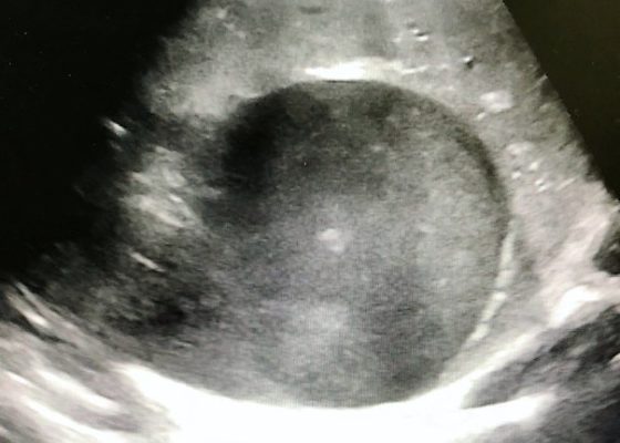
Right Upper Quadrant Pain in a World Explorer
DOI: https://doi.org/10.21980/J8QP9DThe ultrasound images show the abscess, which is a large, circular, hypoechoic mass outlined in blue in the center of the image. The abscess is surrounded by the hyperechoic and heterogeneous liver tissue.
For better delineation of the abscess, a CT was ordered. The axial CT scan image shows the liver abscess, which appears as a hypodense, ovoid, intrahepatic fluid collection within the liver parenchyma. The size of the abscess has been annotated with a dotted line measuring 194.9 mm x 166.2 mm.
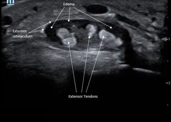
Point-Of-Care Ultrasound for the Diagnosis of Extensor Tenosynovitis
DOI: https://doi.org/10.21980/J8Q050Point-of-care ultrasound of the dorsal aspect of the left hand reveals a heterogenous hypoechoic fluid collection surrounding the extensor tendons (axial view) within the retinaculum consistent with edema. Longitudinal view shows anechoic fluid within the tenosynovium which is located between the anisotropic extensor tendon and linear hyperechoic synovial sheath. Longitudinal view also shows some cobblestoning, or tissue edema, superficial to the anisotropic extensor tendon. The patient’s contralateral right dorsal hand was scanned in a longitudinal view and shows no cobblestoning or hypoechoic fluid under the synovial sheath. The patient was diagnosed with tenosynovitis, and started on intravenous antibiotics.
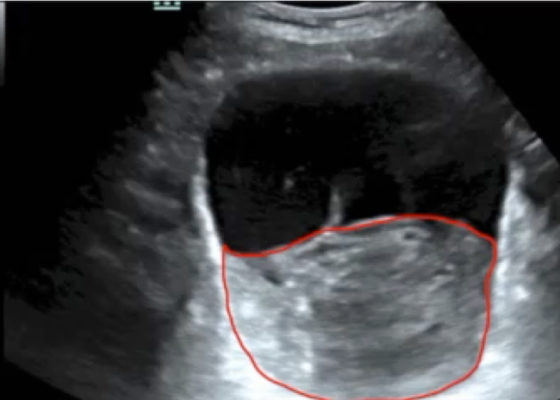
Point-of-care Ultrasound in the Diagnosis and Monitoring of Bladder Hematoma vs. Hemorrhage
DOI: https://doi.org/10.21980/J8092FBladder POCUS demonstrated 500mL of post void residual fluid, indicative of retention. Half of the volume is hyperechoic (red circle); this is likely the bladder wall hematoma. Could also consider sonographic artifact, bladder mass, or cystitis.1-2

Point-of-Care Ultrasound to Evaluate Intrahepatic Biliary Stent Function
DOI: https://doi.org/10.21980/J86S6NThe ultrasound image demonstrates severe intrahepatic biliary ductal dilatation without an obvious intrahepatic obstructive lesion, as pointed out by the white arrows. The hepatic vasculature is well-distinguished from the biliary tree via color flow doppler, as seen by the white arrowheads.

Diagnosis and Treatment of an Anterior Shoulder Dislocation with Bedside Ultrasound
DOI: https://doi.org/10.21980/J8Z924Bedside ultrasound with the transducer placed on the posterior right shoulder revealed an anterior dislocation of the right humerus. This is evident by displacement of the humeral head further away from the posteriorly placed ultrasound transducer, and appears deep to the glenoid cavity. In a posterior shoulder dislocation, the humeral head would appear closer to the transducer (and the near field of the ultrasound image) than the glenoid. Note that a hypoechoic, heterogeneous fluid collection is within the joint space, compatible with a hematoma. A right shoulder X-ray confirmed the anterior dislocation with no evidence of fracture. Under direct ultrasound guidance the glenohumeral joint space was injected with 10 mL of 2% lidocaine as an intraarticular anesthetic block. The right shoulder was reduced using continual traction. Post-reduction ultrasound demonstrated a successful shoulder reduction, depicted by the humeral head being relocated to its anatomical location, adjacent to the glenoid cavity, as noted on the ultrasound image. A hematoma remains present within the joint space. Successful shoulder reduction was further confirmed by X-ray. The patient’s arm was placed in a sling and she was discharged home with orthopedics follow-up.
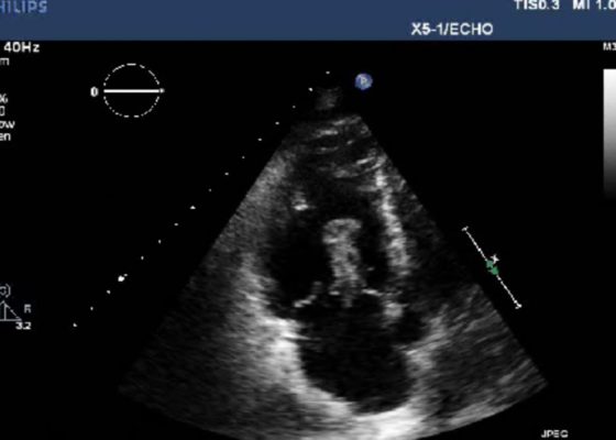
Right Atrial Thrombus
DOI: https://doi.org/10.21980/J8F93V History of present illness: A 77-year-old male presented to the emergency department with shortness of breath. Symptoms progressively worsened over the last 4-5 days, and on arrival was associated with chest tightness. He denied any medical conditions, smoking, or pertinent family history. He has not seen a primary care physician in “many years.” Upon arrival he was in mild