
CT


Pulmonary Arteriovenous Malformation in a Patient with Suspected Hereditary Hemorrhagic Telangiectasia: A Case Report
DOI: https://doi.org/10.21980/J8M353Initial vital signs were unremarkable, including oxygen saturation of 98% on room air. The patient did not exhibit any signs of respiratory distress, and the lungs were clear to auscultation bilaterally. Labs were obtained, which showed normal hemoglobin at 15.8. Computed tomography (CT) of the chest (Video 1) showed a large left upper lobe arteriovenous malformation (AVM) with large feeding arteries and tortuous dilated draining veins (red arrow) measuring up to 3.8cm. Imaging also demonstrated nonspecific multifocal ground-glass opacities, which may have represented pulmonary hemorrhage (blue outline) from AVM without evidence of contrast extravasation to suggest active bleeding.
Case Report: Not Your Typical Kidney Stone
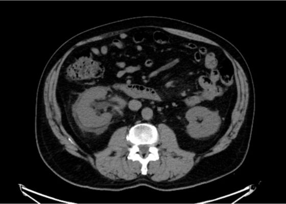
DOI: https://doi.org/10.21980/J8GD2TThe CT scan demonstrates nephrolithiasis with associated forniceal rupture. Encircled in the yellow outline is fluid, demonstrating a forniceal rupture. The stone is in the proximal aspect of the ureter, as highlighted by the purple arrow.
A Case Report of a Transected Carotid Artery Caused by a Stab Wound to the Neck
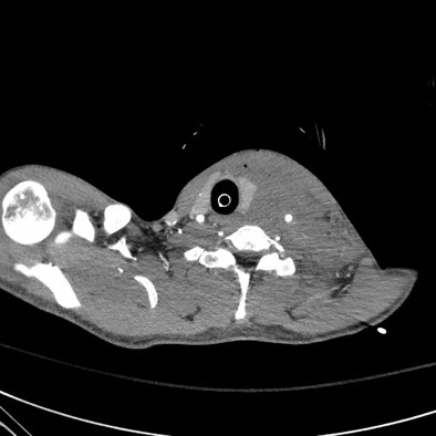
DOI: https://doi.org/10.21980/J8BP8MThe post intubation chest x-ray (CXR) showed severe rightward displacement of the trachea (purple arrow). The computed tomography angiogram (CTA) showed transection of the left common carotid artery (LCCA), extensive neck hematoma without extravasation and severe tracheal deviation to the right (blue arrow). The intravenous (IV) contrasted chest computed tomography (CT) image showed a lateral contrast projection from the aortic arch at the level of the isthmus (green and pink arrows). There were no other significant injuries reported on the CT scans of the chest, abdomen and pelvis.

Posterior Sternoclavicular Dislocation: A Case Report
DOI: https://doi.org/10.21980/J8363QChest X-ray revealed an inferiorly displaced right clavicle at the right sternoclavicular joint (blue arrow). A computed tomography angiogram (CTA) of the chest was therefore obtained and revealed a right posterior sternoclavicular dislocation with resultant compression of the left brachiocephalic vein (purple arrow). Even though the right clavicle is displaced, the anatomy of the brachiocephalic vein is such that it is positioned to the right of midline, placing the left brachiocephalic vein posterior to the right clavicle. The right brachiocephalic and common carotid artery were normal in appearance. The CTA also revealed a comminuted fracture of the left anterior second rib at the costochondral junction that had not been previously seen on the x-ray.
Necrotizing Fasciitis and Mediastinitis after Wisdom Tooth Extraction: A Case Report
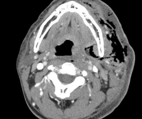
DOI: https://doi.org/10.21980/J8XW7KComputer tomography (CT) imaging of soft tissues of the neck and of the chest/abdomen/pelvis revealed extensive swelling and subcutaneous air (see red arrows) on the left side of the face and neck extending to the left shoulder, as well as parapharyngeal/retropharyngeal spaces and posterior/superior mediastinum.

Adult Clavicular Fracture Case Report
DOI: https://doi.org/10.21980/J8FM0TThe patient's chest and clavicular radiographs showed a comminuted displaced acute fracture of the right mid-clavicle (green, blue, yellow). The clavicular fracture was also visible on the chest computed tomography (CT). The remainder of his trauma workup was negative for acute findings.

A Case Report of Ogilvie’s Syndrome in a 58-year-old Quadriplegic
DOI: https://doi.org/10.21980/J82922Plain radiograph of the patient's abdomen revealed a gaseous distention of the colon. This is demonstrated as noted in the abdominal x-ray as gaseous distention, most notably in the large bowel (arrows) including the rectal region (large circle). Follow up computed tomography (CT) scan affirmed severe pancolonic gaseous distention measuring up to 11.2 cm, compatible with colonic pseudo-obstruction as noted by the large red arrows. No anatomical lesion or mechanical obstruction was observed, as well as no evidence of malignancy or other acute process.
Cecal Volvulus Diagnosed with a Whirl Sign: A Case Report
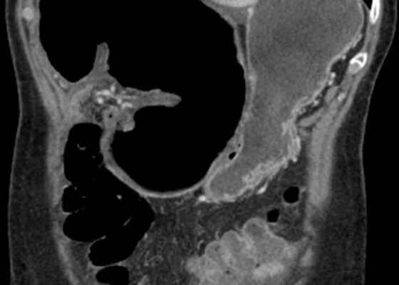
DOI: https://doi.org/10.21980/J8XM05The CT image demonstrates a “whirl sign” (red arrow) which is indicative of a volvulus. This image occurs when bowel, mesentery and vasculature rotate around a transition point causing an image similar to a hurricane on a weather map. When seen on a CT scan, a whirl sign suggests a high likelihood of either a closed loop bowel obstruction or volvulus in the cecum, sigmoid or midgut. In any of the cases, seeing a whirl sign strongly increases the need for emergent surgical management.