
Latest Articles


Pulmonary Arteriovenous Malformation in a Patient with Suspected Hereditary Hemorrhagic Telangiectasia: A Case Report
DOI: https://doi.org/10.21980/J8M353Initial vital signs were unremarkable, including oxygen saturation of 98% on room air. The patient did not exhibit any signs of respiratory distress, and the lungs were clear to auscultation bilaterally. Labs were obtained, which showed normal hemoglobin at 15.8. Computed tomography (CT) of the chest (Video 1) showed a large left upper lobe arteriovenous malformation (AVM) with large feeding arteries and tortuous dilated draining veins (red arrow) measuring up to 3.8cm. Imaging also demonstrated nonspecific multifocal ground-glass opacities, which may have represented pulmonary hemorrhage (blue outline) from AVM without evidence of contrast extravasation to suggest active bleeding.
Case Report: Not Your Typical Kidney Stone
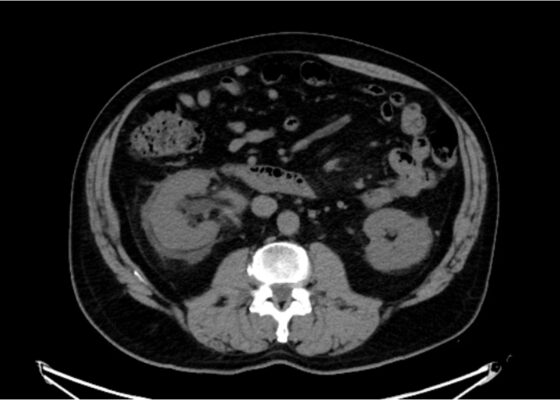
DOI: https://doi.org/10.21980/J8GD2TThe CT scan demonstrates nephrolithiasis with associated forniceal rupture. Encircled in the yellow outline is fluid, demonstrating a forniceal rupture. The stone is in the proximal aspect of the ureter, as highlighted by the purple arrow.
A Case Report of a Transected Carotid Artery Caused by a Stab Wound to the Neck
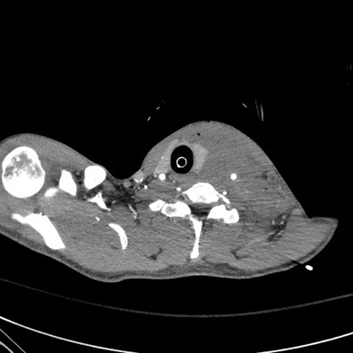
DOI: https://doi.org/10.21980/J8BP8MThe post intubation chest x-ray (CXR) showed severe rightward displacement of the trachea (purple arrow). The computed tomography angiogram (CTA) showed transection of the left common carotid artery (LCCA), extensive neck hematoma without extravasation and severe tracheal deviation to the right (blue arrow). The intravenous (IV) contrasted chest computed tomography (CT) image showed a lateral contrast projection from the aortic arch at the level of the isthmus (green and pink arrows). There were no other significant injuries reported on the CT scans of the chest, abdomen and pelvis.
Case Report of COVID-19 Positive Male with Late-Onset Full Body Maculopapular Rash
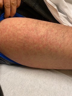
DOI: https://doi.org/10.21980/J86W72The images demonstrate a diffuse, flat, maculopapular exanthema along the torso, bilateral upper and lower extremities, and neck without edema consistent with reported cutaneous manifestations of COVID-19. There are no surrounding bullae, vesicles, or draining. On palpation, there was blanching of the rash. Sensation to light touch was intact in all extremities. The findings were also apparent on the face with no mucosal involvement.

Posterior Sternoclavicular Dislocation: A Case Report
DOI: https://doi.org/10.21980/J8363QChest X-ray revealed an inferiorly displaced right clavicle at the right sternoclavicular joint (blue arrow). A computed tomography angiogram (CTA) of the chest was therefore obtained and revealed a right posterior sternoclavicular dislocation with resultant compression of the left brachiocephalic vein (purple arrow). Even though the right clavicle is displaced, the anatomy of the brachiocephalic vein is such that it is positioned to the right of midline, placing the left brachiocephalic vein posterior to the right clavicle. The right brachiocephalic and common carotid artery were normal in appearance. The CTA also revealed a comminuted fracture of the left anterior second rib at the costochondral junction that had not been previously seen on the x-ray.
Case Report: Traumatic Tension Pneumothorax in a Pediatric Patient
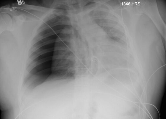
DOI: https://doi.org/10.21980/J8ZD1SChest X-ray demonstrated significant right-sided pneumothorax (with red outline showing border of collapsed right lung) with cardio mediastinal shift to the left (shown by blue arrows) indicative of a tension pneumothorax

Vomiting in Pediatric Patients
DOI: https://doi.org/10.21980/J8P363By the end of this TBL session, learners should be able to: 1) Identify red flag symptoms that should prompt referral for urgent intervention by GI or surgical specialists; 2) recognize how chronicity of the vomiting can alter the differential diagnosis; 3) describe the varying pathways that can cause nausea and vomiting; 4) determine the necessity of imaging tests to confirm and possibly treat various causes of vomiting; 5) interpret imaging studies associated with specific causes of vomiting.
Febrile Seizure Team-based Learning
DOI: https://doi.org/10.21980/J8JD12By the end of this educational session, the learner will: 1) list the characteristics of a simple febrile seizure; 2) discuss the management of a child with a simple vs. complex febrile seizure; 3) discuss the risk factors that correlate with an increased risk of a subsequent febrile seizure; 4) determine when a lumbar puncture should be considered in a febrile child with a seizure; 5) identify when to give anti-epileptics and construct an algorithm for their use; 6) discuss with parents, provide education and return precautions.

What do you do if your relief comes to work intoxicated: An Impaired Provider Scenario
DOI: https://doi.org/10.21980/J8DM0HBy the end of this simulation, learners will be able to: 1) Identify potential impairment in the form of alcohol intoxication in a physician colleague; 2) demonstrate the ability to communicate effectively with the colleague and remove them from the patient care environment; 3) discuss the appropriate next steps in identifying long-term wellness resources for the impaired colleague; and 4) demonstrate understanding of the need to continue to provide care for the patients by moving the case forward.
Extracorporeal Membrane Oxygenation (ECMO) for Refractory Cardiac Arrest
DOI: https://doi.org/10.21980/J88W69ABSTRACT: Audience: Our target audience includes emergency medicine residents/physicians. Introduction: Treating cardiac arrest is a common theme during simulated emergency medicine training; however, less time is focused on treating refractory cases of cardiac arrest. There are varying definitions of refractory cardiac arrest, but it is most commonly defined as the inability to obtain return of spontaneous circulation (ROSC) after 10-30