Visual EM
Achalasia: An Uncommon Presentation with Classic Imaging
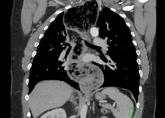
DOI: https://doi.org/10.21980/J86D2BThe chest X-ray demonstrated a markedly widened mediastinum (red brackets), raising concern for thoracic aortic aneurysm/aortic dissection, which prompted labs and contrast-enhanced computed tomography (CT) of the chest. The CT revealed a dilated proximal esophagus that narrowed distally (yellow tracing and red arrow), with particulate material, mass-effect on the trachea (purple outline), and bilateral patchy opacities suggesting aspiration. Barium esophagram showed a drastically dilated esophagus filled with contrast (yellow arrow), terminating into the classic “bird’s beak sign” (red arrow) at the lower esophageal sphincter (LES). Esophageal manometry later confirmed achalasia, proving that widened mediastina can have unexpected etiologies.
Right Ventricular Dilation in Patient With Submassive Pulmonary Embolism
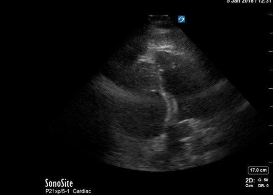
DOI: https://doi.org/10.21980/J82P84Bedside echocardiography four chamber view revealed enlarged right ventricular (RV) to left ventricular (LV) ratio (greater than 1) on apical four-chamber view (see red and blue outlines respectively). The right atrium is not clearly delineated in this image and therefore is not outlined. One can also rule out a large pericardial effusion as the cause of her dyspnea, since there is no large hypoechoic collection surrounding the heart on either four- chamber view or parasternal long view.
Traumatic Hyphema
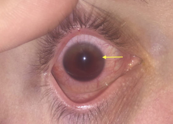
DOI: https://doi.org/10.21980/J8Z04SUpon initial evaluation, the patient had an obvious hyphema in the right eye with associated conjunctival injection. Initially, the bleeding in the anterior chamber was cloudy just above the level of the pupil (yellow arrow), appearing to possibly be a grade II hyphema. There were no other signs of trauma to the eye under Wood’s lamp examination with fluorescein staining. The globe was intact. Intraocular pressure in the affected eye was 19 mmHg and 15 mmHg in the unaffected eye. Extraocular movements were full and intact. The pupil was 4 mm round and reactive to direct and consensual light. Visual acuity was greater than 20/200 in the affected eye compared to 20/25 in the unaffected eye. After an observation period of two hours, with the patient remaining upright, the hyphema had settled down to a rim in the lower anterior chamber (green arrow), a grade I hyphema.
Point of Care Ultrasound Illustrating Small Bowel Obstruction
DOI: https://doi.org/10.21980/J8T637POCUS of the small bowel illustrated significantly dilated loops of bowel (white line), thickened bowel wall (white arrow) and to-and-fro peristalsis, consistent with small bowel obstruction.
An Unusual Case of Pharyngitis: Herpes Zoster of Cranial Nerves 9, 10, C2, C3 Mimicking a Tumor
DOI: https://doi.org/10.21980/J8B05KOn exam, the patient was sitting upright while holding an emesis basin filled with saliva. His voice was noticeably hoarse. Examination of the head and neck revealed vesicular eruptions on the left scalp in the V1 dermatome and on the left mastoid process (Images 1 and 2). Physical exam also shows vesicular eruptions on the left posterior oropharynx that did not cross midline (Image 3).
Ramsay Hunt Syndrome
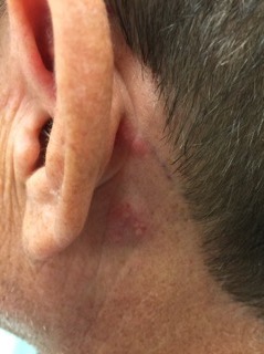
DOI: https://doi.org/10.21980/J85S7QLeft-sided cranial nerve VII palsy with flattened forehead creases, inability to keep the left eye open, and drooping of the corner of mouth. Vesicular lesions were found in and posterior to the left ear in a unilateral, dermatomal distribution.
Retinal Detachment
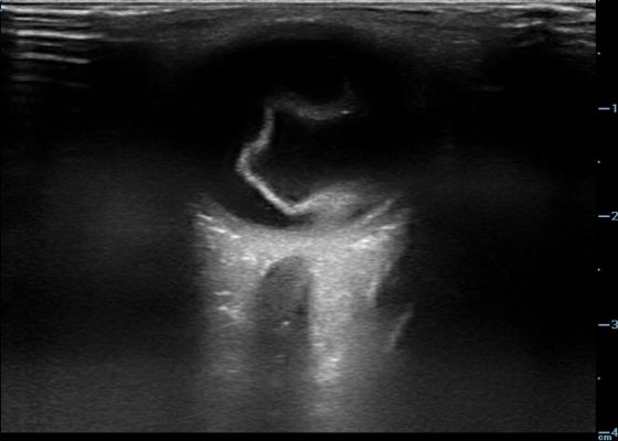
DOI: https://doi.org/10.21980/J8204QBedside ocular ultrasound revealed a serpentine, hyperechoic membrane that appeared tethered to the optic disc posteriorly with hyperechoic material underneath. These findings are consistent with retinal detachment (RD) and associated retinal hemorrhage.
Necrotizing Soft Tissue Infection
DOI: https://doi.org/10.21980/J8X92TComputed tomography (CT) of the abdominal and pelvis with intravenous (IV) contrast revealed inflammatory changes, including gas and fluid collections within the ventral abdominal wall extending to the vulva, consistent with a necrotizing soft tissue infection.

