
JETem
  • Home
  • About
    • Aim and Scope
    • Our Team
    • Editorial Board
    • FAQ
  • Issues
    • Current Issue
    • Ahead of Print
    • Past Issues
  • Visual EM
    • Latest Visual EM
    • Search Visual EM
    • Thumbnail Library
  • For Authors
    • Instructions for Authors
    • Submit to JETem
    • Photo Consent
    • Policies
      • Peer Review Policy
      • Copyright Policy
      • Editorial Policy, Ethics and Responsibilities
      • Conflicts of Interest & Informed Consent
      • Open Access Policy
      • Acceptance Fee
  • For Reviewers
    • Instructions for JETem Reviewers
    • Interested in Being a JETem Reviewer?
  • Topic
    • Abdominal/Gastroenterology
    • Administration
    • Board Review
    • Cardiology/Vascular
    • Certifying Exam Cases
      • Clinical Decision-Making
      • Prioritization
      • Communication
      • Procedure & Ultrasound
    • Clinical Informatics, Telehealth and Technology
    • Dermatology
    • EMS
    • Endocrine
    • ENT
    • Faculty Development
    • Genitourinary
    • Geriatrics
    • Hematology/Oncology
    • Infectious Disease
    • Miscellaneous (stats, etc)
    • Neurology
    • Ob/Gyn
    • Ophthalmology
    • Orthopedics
    • Pediatrics
    • Pharmacology
    • Procedures
    • Psychiatry
    • Renal/Electrolytes/Nutrition
    • Respiratory
    • Social Determinants of Health
    • Toxicology
    • Trauma
    • Ultrasound
    • Urology
    • Wellness
    • Wilderness
  • Modality
    • Certifying Exam Practice Cases
      • Clinical Care Cases
      • Communication & Procedural Cases
      • Old School Oral Board Cases
    • Curricula
    • Innovations
    • Lectures
    • Podcasts
    • Simulation
    • Small Group Learning
    • Team Based Learning (TBL)
    • Visual EM
  • Contact Us

Visual EM

Creative Commons images

FAST Exam to Diagnose Subcapsular Renal Hematoma

Michelle Sofia Bakardjiev, MD* and Amanda Esposito, MD^

DOI: https://doi.org/10.21980/J8NP8DIssue 4:4 [mrp_rating_result]
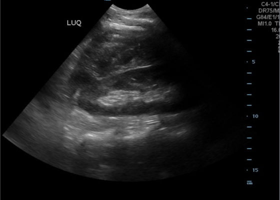
A bedside point of care ultrasound FAST exam was performed revealing a left subcapsular renal hematoma. The hematoma was a non-compressing hematoma, evidenced by preserved renal contour with the hematoma labeled with a red H and the normal renal contour labeled with a green K. Additionally, cortical necrosis and ischemia can be characterized by a dark, hypoechogenic renal cortex on ultrasonography with a decrease in flow to the cortex on color doppler which was not seen on this patient, providing further evidence that the hematoma was non-compressing. The hematoma was concluded to be an acute process due to its hypoechoic appearance with some mixed ultrasonographic echoes caused by the early deposit of fibrin.
Renal/ElectrolytesVisual EM
Creative Commons images

Right Upper Quadrant Pain in a World Explorer

Julia Hutchison, DO* and Nur-Ain Nadir, MD, MHPE*

DOI: https://doi.org/10.21980/J8QP9D Issue 4:4 [mrp_rating_result]
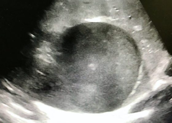
The ultrasound images show the abscess, which is a large, circular, hypoechoic mass outlined in blue in the center of the image. The abscess is surrounded by the hyperechoic and heterogeneous liver tissue. For better delineation of the abscess, a CT was ordered. The axial CT scan image shows the liver abscess, which appears as a hypodense, ovoid, intrahepatic fluid collection within the liver parenchyma. The size of the abscess has been annotated with a dotted line measuring 194.9 mm x 166.2 mm.
Abdominal/GastroenterologyVisual EM
Creative Commons images

Point-Of-Care Ultrasound for the Diagnosis of Extensor Tenosynovitis

James A Frank, MD*, Joshua Lupton, MD* and Bryson Hicks, MD*

DOI: https://doi.org/10.21980/J8Q050Issue 4:3[mrp_rating_result]
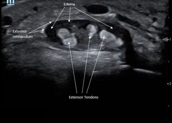
Point-of-care ultrasound of the dorsal aspect of the left hand reveals a heterogenous hypoechoic fluid collection surrounding the extensor tendons (axial view) within the retinaculum consistent with edema. Longitudinal view shows anechoic fluid within the tenosynovium which is located between the anisotropic extensor tendon and linear hyperechoic synovial sheath. Longitudinal view also shows some cobblestoning, or tissue edema, superficial to the anisotropic extensor tendon. The patient’s contralateral right dorsal hand was scanned in a longitudinal view and shows no cobblestoning or hypoechoic fluid under the synovial sheath. The patient was diagnosed with tenosynovitis, and started on intravenous antibiotics.
OrthopedicsInfectious DiseaseUltrasoundVisual EM
Creative Commons images

Point-of-care Ultrasound in the Diagnosis and Monitoring of Bladder Hematoma vs. Hemorrhage

Esther Kim, BS* and Shadi Lahham, MD, MS*

DOI: https://doi.org/10.21980/J8092FIssue 4:3[mrp_rating_result]
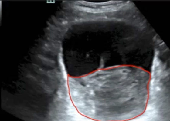
Bladder POCUS demonstrated 500mL of post void residual fluid, indicative of retention. Half of the volume is hyperechoic (red circle); this is likely the bladder wall hematoma. Could also consider sonographic artifact, bladder mass, or cystitis.1-2
GenitourinaryUltrasoundVisual EM
Creative Commons images

Asymptomatic CT Iodinated Contrast Extravasation of the Upper Extremity

Eric Liao, MD* and John Costumbrado, MD, MPH*

DOI: https://doi.org/10.21980/J8VK87 Issue 4:3[mrp_rating_result]
The two radiographs demonstrate extravasation of radiopaque iodinated contrast in the lower left upper extremity with most seen in the left antecubital fossa and left proximal forearm. Extravasation is seen in the subcutaneous and subfascial tissue.
Visual EMOrthopedics
Creative Commons images

Oral Herpes Zoster

Daniel Polvino, MD*, Grant Wei, MD* and Christopher Bryczkowski, MD*

DOI: https://doi.org/10.21980/J8QS69Issue 4:3[mrp_rating_result]
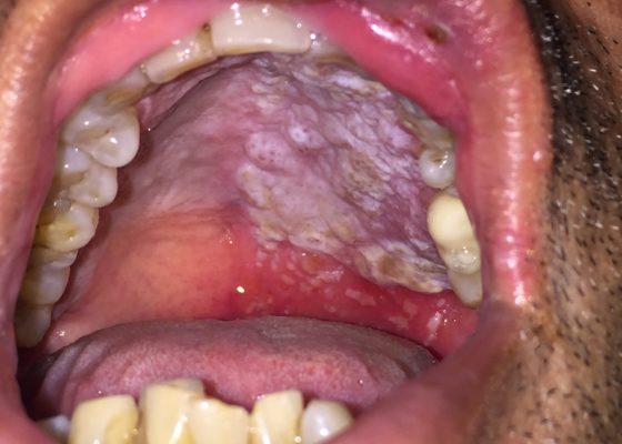
Physical exam findings revealed vesicular lesions on the lip, hard and soft palates which did not cross the midline. The lesions appeared in the distribution of the maxillary branch (V2) of the trigeminal nerve, consistent with herpes zoster.
DermatologyInfectious DiseaseVisual EM
Creative Commons images

Ruptured AAA Presenting with Unresponsiveness and Cardiac Arrest

Lana S Shaker, MD*, Michael W Ullo, MD* and Aislinn Black, DO, MPH*

DOI: https://doi.org/10.21980/J8M34QIssue 4:3[mrp_rating_result]
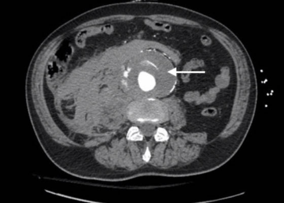
Axial CT images from the CT chest, abdomen and pelvis revealed a large infra-renal abdominal aortic aneurysm measuring 7.3 x 8.2 x 10 cm with extensive mural thrombus (single white arrow) that has ruptured, with active extravasation (black arrow) of contrast into the aneurysm sac and retroperitoneum with large right retroperitoneal hematoma (multiple white arrows).
Cardiology/VascularVisual EM
Creative Commons images

Spontaneous Intracranial Hemorrhage in Severe Hemophilia A: A Rare Cause of Seizure in a Young Child

Sharon Kook Won, MD* and Andrea T Cruz, MD, MPH*

DOI: https://doi.org/10.21980/J8G91D Issue 4:3[mrp_rating_result]
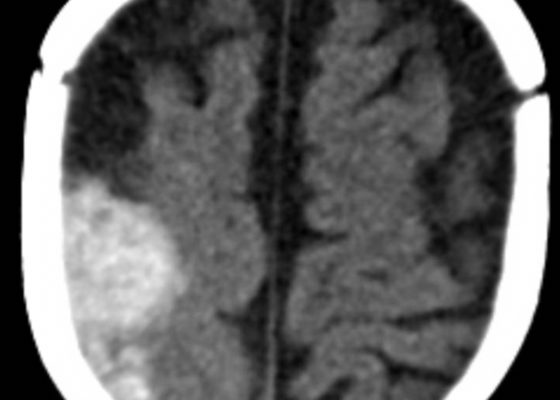
A computed tomography (CT) scan of the head without contrast was obtained out of concern for intracranial pathology due to the patient’s young age and the witnessed focal seizure.  The CT showed a 4.2 x 1.2 x 1.5 cm acute extra-axial intracranial right frontoparietal hemorrhage favoring epidural over subdural hemorrhage given its lenticular shape. There was no underlying fracture, herniation or midline shift identified.
Hematology/OncologyNeurologyVisual EM
«‹21›»
Page 21 of 47

JETem is an online, open access, peer-reviewed, journal-repository for EM educators. We are PMC Indexed.

Most Viewed

  • Simple Auricular Hematoma (“Cauliflower Ear”) Task Trainer
  • The Three Breath Method: Teaching Coping Mechanisms for Processing Patient Death
  • Clinical Decision Making: Acute Ischemic Stroke with Large Vessel Occlusion
  • PRIMER: Premedical Introduction to Mentored Emergency Research: Connecting Undergraduate Assistants and Emergency Medicine Residents in a Community Hospital Setting
  • Dangerous Dysrhythmias in the ED: An Interactive EKG Workshop for Emergency Medicine Clerkship Learners

Visit Our Collaborators


Creative Commons Licence
This work is licensed under a Creative Commons Attribution 4.0 International License.

About

Education

Learners should benefit from active learning. JETem accepts submissions of team-based learning, small group learning, simulation, podcasts, lectures, innovations, curricula, question sets, and visualEM.

Scholarship

We believe educators should advance through the scholarship of their educational work. JETem gives educators the opportunity to publish scholarly academic work so that it may be widely distributed, thereby increasing the significance of their results.

Links

  • Home
  • Aim and Scope
  • Current Issue
  • For Reviewers
  • Instructions for Authors
  • Contact Us

Newsletter

Sign up to receive updates from JETem regarding newly published issues and findings.

Copyright Creative Commons Attribution 4.0 International