
Ultrasound
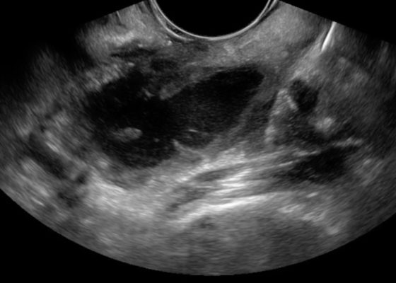

Bedside Ultrasound for the Diagnosis of Peritonsillar Abscess
DOI: https://doi.org/10.21980/J8N33NThe first video is an intraoral ultrasound using the high frequency endocavitary probe demonstrating an anechoic fluid collection adjacent to the patient’s enlarged left tonsil. The second video shows real-time ultrasound-guided successful drainage of the PTA.
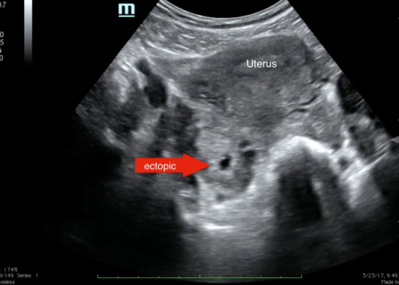
Point-of-care Ultrasound for the Diagnosis of Ectopic Pregnancy
DOI: https://doi.org/10.21980/J8VK7VThe transabdominal pelvic ultrasound shows an empty uterus (annotated) with free fluid and a right sided extrauterine gestational sac representing an ectopic pregnancy (red arrow).


Introducing point-of-care ultrasound through competency-based simulation education using a fractured chicken bone model
DOI: https://doi.org/10.21980/J8GG95To introduce medical students to PoCUS with an inexpensive, reproducible, and educationally effective model using fractured chicken bones set in gelatin, and to assess medical students’ abilities to identify simulated long-bone fractures using PoCUS.
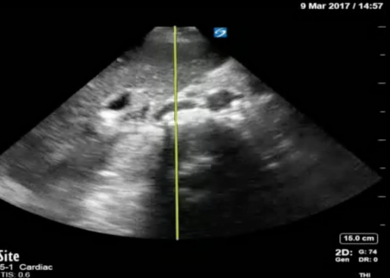
Using Bedside Ultrasound to Rapidly Differentiate Shock
DOI: https://doi.org/10.21980/J8S047A RUSH exam demonstrated hyperdynamic cardiac contractility and collapse of the inferior vena cava (IVC) with probe compression more than 50% suggesting hypovolemia likely secondary to sepsis. Incidentally, Morrison’s pouch revealed a large right renal cyst but no signs of free fluid. A computed tomography of abdomen/pelvis showed a 10.8 x 9.5 cm right renal cyst and left lower lobe pneumonia.
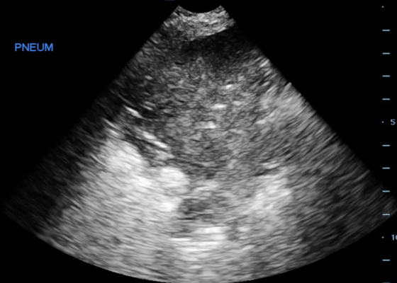
Pneumonia Diagnosed by Point-of-Care Ultrasound
DOI: https://doi.org/10.21980/J8XP64Point-of-care ultrasound of the left lower lobe demonstrates lung hepatization, a classic finding for pneumonia. In addition, a shred sign is present with both air bronchograms and focal B-lines—all suggestive of poorly aerated, consolidated lung. The patient was started on antibiotics and admitted to the hospital with a diagnosis of community-acquired pneumonia.
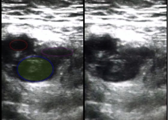
Use of Bedside Compression Ultrasonography for Diagnosis of Deep Venous Thrombosis
DOI: https://doi.org/10.21980/J81G94As shown in the still image of the performed ultrasound, a transverse view of the proximal-thigh revealed a visible thrombus (green shading) occluding the lumen of the left common femoral vein (blue ring), which was non-compressible when direct pressure was applied to the probe. Also visible is a patent and compressible branch of the common femoral vein (purple ring) and the femoral artery (red ring), highlighted by its thick vessel wall and pulsatile motion.

Emed-Opoly: Echocardiography
DOI: https://doi.org/10.21980/J8PC77By the end of this session, the learner will be able to:
1) Recognize normal and abnormal left heart global function
2) Recognize normal and abnormal right heart global function
3) Recognize pericardial effusions and pericardial tamponade

A Toddler with Abdominal Pain and Emesis
DOI: https://doi.org/10.21980/J8XW2PIn the long axis video, the appendix appears as an enlarged, non-compressible, blind-ending tubular structure (white arrow) with distinct appendiceal wall layers and lack of peristalsis. In the short axis video, the appendix appears as a target sign (yellow arrow) between the abdominal and psoas muscles. The maximal outer diameter (MOD) measures 11.8mm and the appendix wall measures 0.17mm. There is trace adjacent free fluid and echogenic periappendiceal fat. Transverse axis video and image (red arrow) demonstrate that the appendix is not compressible. These findings are consistent with acute appendicitis.