
JETem
  • Home
  • About
    • Aim and Scope
    • Our Team
    • Editorial Board
    • FAQ
  • Issues
    • Current Issue
    • Ahead of Print
    • Past Issues
  • Visual EM
    • Latest Visual EM
    • Search Visual EM
    • Thumbnail Library
  • For Authors
    • Instructions for Authors
    • Submit to JETem
    • Photo Consent
    • Policies
      • Peer Review Policy
      • Copyright Policy
      • Editorial Policy, Ethics and Responsibilities
      • Conflicts of Interest & Informed Consent
      • Open Access Policy
      • Acceptance Fee
  • For Reviewers
    • Instructions for JETem Reviewers
    • Interested in Being a JETem Reviewer?
  • Topic
    • Abdominal/Gastroenterology
    • Administration
    • Board Review
    • Cardiology/Vascular
    • Certifying Exam Cases
      • Clinical Decision-Making
      • Prioritization
      • Communication
      • Procedure & Ultrasound
    • Clinical Informatics, Telehealth and Technology
    • Dermatology
    • EMS
    • Endocrine
    • ENT
    • Faculty Development
    • Genitourinary
    • Geriatrics
    • Hematology/Oncology
    • Infectious Disease
    • Miscellaneous (stats, etc)
    • Neurology
    • Ob/Gyn
    • Ophthalmology
    • Orthopedics
    • Pediatrics
    • Pharmacology
    • Procedures
    • Psychiatry
    • Renal/Electrolytes/Nutrition
    • Respiratory
    • Social Determinants of Health
    • Toxicology
    • Trauma
    • Ultrasound
    • Urology
    • Wellness
    • Wilderness
  • Modality
    • Certifying Exam Practice Cases
      • Clinical Care Cases
      • Communication & Procedural Cases
      • Old School Oral Board Cases
    • Curricula
    • Innovations
    • Lectures
    • Podcasts
    • Simulation
    • Small Group Learning
    • Team Based Learning (TBL)
    • Visual EM
  • Contact Us

Trauma

Creative Commons images

Posterior Sternoclavicular Dislocation: A Case Report

Stephanie Songey, DO*, Christopher Goodwill, DO* and Kimberly Sokol, MD, MS, MACM*

DOI: https://doi.org/10.21980/J8363Q Issue 6:1[mrp_rating_result]
Chest X-ray revealed an inferiorly displaced right clavicle at the right sternoclavicular joint (blue arrow). A computed tomography angiogram (CTA) of the chest was therefore obtained and revealed a right posterior sternoclavicular dislocation with resultant compression of the left brachiocephalic vein (purple arrow). Even though the right clavicle is displaced, the anatomy of the brachiocephalic vein is such that it is positioned to the right of midline, placing the left brachiocephalic vein posterior to the right clavicle. The right brachiocephalic and common carotid artery were normal in appearance. The CTA also revealed a comminuted fracture of the left anterior second rib at the costochondral junction that had not been previously seen on the x-ray.
OrthopedicsTraumaVisual EM
Creative Commons images

Case Report: Traumatic Tension Pneumothorax in a Pediatric Patient

Zachary Tritsch, MD*, Gayle Galan, MD*, Gary Oates, MD* and Janelle Thomas, MD*

DOI: https://doi.org/10.21980/J8ZD1S Issue 6:1[mrp_rating_result]
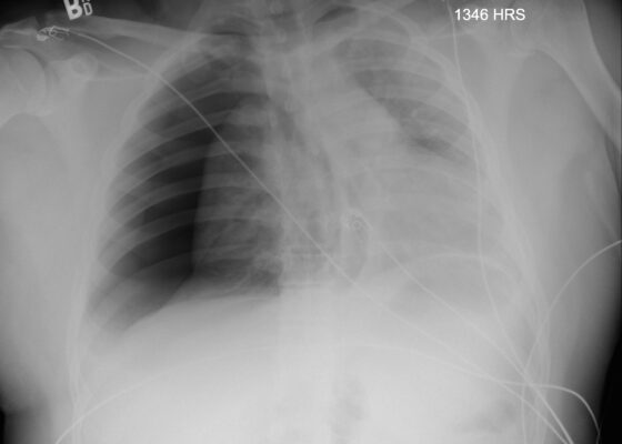
Chest X-ray demonstrated significant right-sided pneumothorax (with red outline showing border of collapsed right lung) with cardio mediastinal shift to the left (shown by blue arrows) indicative of a tension pneumothorax
TraumaPediatricsRespiratoryVisual EM

Owning the Trauma Bay: Teaching Trauma Resuscitation to Emergency Medicine Residents and Nurses through In-situ Simulation

Andrew Bellino, MD*, Alexandra June Gordon, MD^, Al’ai Alvarez, MD*  and Kimberly Schertzer, MD*

DOI: https://doi.org/10.21980/J8WK9X Issue 5:4[mrp_rating_result]
ABSTRACT: Audience: The following two cases were designed to address learning objectives specific to interns, junior residents, and senior residents in emergency medicine, as well as trauma-certified emergency nurses.  Introduction: Traumatic and unintentional injuries account for 5.8 million deaths across the globe each year, with a high proportion of those deaths occurring within the initial hour from the time of
SimulationTrauma

Simulated Mass Casualty Incident Triage Exercise for Training Medical Personnel

Alaina Brinley Rajagopal, MD, PhD*, Nathan Jasperse, BS* and Megan Boysen Osborn, MD, MHPE* 

DOI: https://doi.org/10.21980/J82H1R Issue 5:4[mrp_rating_result]
By the end of this exercise, learners should be able to (1) recite the basic START patient categories (2) discuss the physical exam signs associated with each START category, (3) assign roles to medical providers in a mass casualty scenario, (4) accurately categorize patients into triage categories: green, yellow, red, and black, and (5) manage limited resources when demand exceeds availability.
EMSSmall Group LearningTrauma
Creative Commons images

Adult Clavicular Fracture Case Report

Jessica L Sea, PhD*, Nadia Zuabi, MD* and Alisa Wray, MD, MAEd*

DOI: https://doi.org/10.21980/J8FM0TIssue 5:4[mrp_rating_result]
The patient's chest and clavicular radiographs showed a comminuted displaced acute fracture of the right mid-clavicle (green, blue, yellow). The clavicular fracture was also visible on the chest computed tomography (CT). The remainder of his trauma workup was negative for acute findings.
OrthopedicsTraumaVisual EM
DRUJ Elbow Xray, Lateral. Unannotated. JETem 2020

Case Report of Distal Radioulnar Joint and Posterior Elbow Dislocation

Danielle Matonis, MD*, Katelyn Wittel, BS* and Alisa Wray, MD, MAEd*

DOI: https://doi.org/10.21980/J89S6K Issue 5:4[mrp_rating_result]
Radiographs of the left elbow and wrist were obtained. Left elbow radiographs showed simple posterolateral dislocation of the olecranon (red) without fracture of the olecranon (red) or trochlea (blue). Left wrist lateral radiographs demonstrated DRUJ dislocation with dorsal displacement of the distal ulna (green) without fracture or widening of the radioulnar joint (purple). Post-reduction radiographs demonstrated appropriate alignment of the elbow with the trochlea seated in the olecranon and improved alignment of the DRUJ.
OrthopedicsTraumaVisual EM
Creative Commons images

A Case Report of Epidural Hematoma After Traumatic Brain Injury

Ronald Goubert, BS*, Alisa Wray, MD, MAEd* and Danielle Matonis, MD

DOI: https://doi.org/10.21980/J8R059 Issue 5:3[mrp_rating_result]
Non-contrast CT head demonstrated a right sided EDH (red arrow) with overlying scalp hematoma, left-sided subdural hematoma (blue arrow), and bilateral subarachnoid hemorrhages. No skull fractures were noted.
TraumaNeurologyVisual EM
Creative Commons images

Traumatic Diaphragmatic Rupture – A Case Report

Vincent Hussey, MS*  and Claire Thomas, MD*

DOI: https://doi.org/10.21980/J8G64H Issue 4:4 [mrp_rating_result]
Chest X-ray showed an elevated left hemi-diaphragm with superior displacement of a portion of intra-abdominal contents presumed to be the stomach (green arrowheads) with associated rightward mediastinal shift (yellow arrows). The diagnosis was confirmed by CT. Computed tomography imaging of the chest showed a large, left diaphragmatic defect measuring approximately 5.5 cm with herniation of the upper half of the stomach through the defect. The fundus of the stomach (blue arrow) herniated superiorly through the ruptured diaphragm (red arrow).
TraumaAbdominal/GastroenterologyVisual EM
«‹3›»
Page 3 of 6

JETem is an online, open access, peer-reviewed, journal-repository for EM educators. We are PMC Indexed.

Most Viewed

  • Simple Auricular Hematoma (“Cauliflower Ear”) Task Trainer
  • The Three Breath Method: Teaching Coping Mechanisms for Processing Patient Death
  • Clinical Decision Making: Acute Ischemic Stroke with Large Vessel Occlusion
  • PRIMER: Premedical Introduction to Mentored Emergency Research: Connecting Undergraduate Assistants and Emergency Medicine Residents in a Community Hospital Setting
  • Dangerous Dysrhythmias in the ED: An Interactive EKG Workshop for Emergency Medicine Clerkship Learners

Visit Our Collaborators


Creative Commons Licence
This work is licensed under a Creative Commons Attribution 4.0 International License.

About

Education

Learners should benefit from active learning. JETem accepts submissions of team-based learning, small group learning, simulation, podcasts, lectures, innovations, curricula, question sets, and visualEM.

Scholarship

We believe educators should advance through the scholarship of their educational work. JETem gives educators the opportunity to publish scholarly academic work so that it may be widely distributed, thereby increasing the significance of their results.

Links

  • Home
  • Aim and Scope
  • Current Issue
  • For Reviewers
  • Instructions for Authors
  • Contact Us

Newsletter

Sign up to receive updates from JETem regarding newly published issues and findings.

Copyright Creative Commons Attribution 4.0 International