
Cardiology/Vascular

Torsade de Pointes Due to Hypokalemia and Hypomagnesemia
DOI: https://doi.org/10.21980/J8JP8GBy the end of this simulation session, learners will be able to: 1) formulate appropriate work-up for altered mental status (AMS) 2) recognize hypokalemia and associated findings on ECG 3) address hypomagnesemia in a setting to hypokalemia 4) manage pulseless VT by following advanced cardiac life support (ACLS) 5) recognize and address TdP 6) provide care after return of spontaneous circulation (ROSC) 7) consult intensivist and admit to intensive care unit (ICU).


Inferior STEMI Electrocardiogram in a Young Postpartum Female with Sickle Cell Trait with Chest Pain – A Case Report
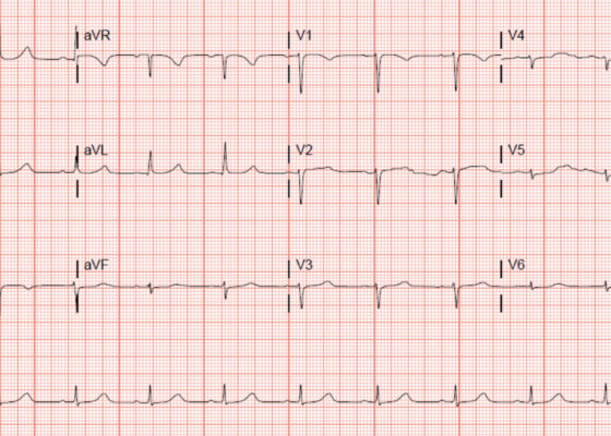
DOI: https://doi.org/10.21980/J8KP95ECG shows evidence of ST segment elevation in the inferolateral leads with reciprocal change in a bigeminy pattern. The ECG pattern seen in this patient demonstrates ST elevations in the inferior leads (II, III, and avF) as well as the precordial leads V4-V6. Reciprocal changes can also be seen in leads I and avL. Though this STEMI pattern is typically associated with occlusion of the right coronary artery in 80% of cases, it may also be caused by occlusion of the left circumflex artery. This may explain this patient’s cardiac catheterization findings of vasospasm in the left circumflex coronary artery.
Aortic Dissection Presenting as a STEMI
DOI: https://doi.org/10.21980/J8W647At the conclusion of the simulation session or during the debriefing session, learners will be able to: 1) Verbalize the anatomical differences and management of Stanford type A and type B aortic dissections, 2) Describe physical exam findings that may be found with ascending aortic dissections, 3) Describe the various clinical manifestations of the propagation of aortic dissections, 4) Discuss the management of aortic dissection, including treatment and disposition.
‘Cath’ It Before It’s Too Late: A Case Report of ECG Abnormalities Indicative of Acute Pathology Requiring Immediate Catheterization
DOI: https://doi.org/10.21980/J8HW7VA 12 lead ECG performed at the time of emergency department (ED) admission revealed regular sinus rhythm with noted T-wave inversion (blue arrows on Figure 1) in Lead aVL new when compared to an ECG performed a few months prior (see Figure 3). Two days later a second ECG was done when the patient developed acute chest pain while in the ICU (Figure 2) that showed persistent inversion in Lead aVL (blue arrows) as well as new J point deviation (JPD) in Leads II, aVF, V5 and V6; and new JPD in Leads V1 and V2 (green arrows) from her previous ECG while in the emergency department. These focal repolarization abnormalities did not qualify as an ST-elevation myocardial infarction by current guidelines.

A Case Report of May-Thurner Syndrome Identified on Abdominal Ultrasound
DOI: https://doi.org/10.21980/J8C64KThe patient initially received a venous doppler ultrasound that showed no evidence of a right or left femoropopliteal venous thrombus. Due to the high suspicion of a DVT given the symmetric swelling to the entire limb and acute onset of pain, a CTV was ordered. The transverse view of the CTV showed chronic thrombotic occlusion of the proximal left common iliac vein associated with compression from the right common iliac artery (figure 1, transverse image of CTA), as well as thrombotic occlusion of the left internal iliac vein tributary and corresponding left ascending lumbar vein. Given the previously mentioned clinical context, these features suggested the diagnosis of May-Thurner syndrome.

Unravelling the Mystery of a Continuous Coil: A Case Report
DOI: https://doi.org/10.21980/J8PM00A CT scan of the abdomen and pelvis with intravenous contrast for evaluation of new onset abdominal pain and distension was obtained in the emergency department. The axial view (CT Image A) shows the coil pack from the prior coil-assisted retrograde transvenous obliteration procedure, seen in the left renal vein and gastric varix (red arrow). The path of the coil (yellow arrow) is continuous into the inferior vena cava (CT Image B). It is then seen (CT Image C) situated in the right ventricle (green arrow). Finally, the coil pack is seen in a coronal section, demonstrating its upward path (blue arrow) in the inferior vena cava. (CT Image D). Additional findings included ascites with advanced cirrhosis. As noted in the CT images, a vascular embolization coil was seen within a varix near the junction of the left renal vein. This appeared to have unraveled and extended superiorly into the inferior vena cava and ultimately into the right atrium and right ventricle.

Principles of Hypotensive Shock: A Video Introduction to Pathophysiology and Treatment Strategies
DOI: https://doi.org/10.21980/J8MS84By the end of this module, participants should be able to: 1) review basic principles of cardiovascular physiology; 2) describe the 4 general pathophysiologic mechanisms of hypotensive shock; 3) recognize various etiologies for each mechanism of hypotensive shock; 4) recognize differences in the clinical presentation of each mechanism of hypotensive shock; 5) cite the basic approach to treatment for each mechanism of hypotensive shock.