
JETem
  • Home
  • About
    • Aim and Scope
    • Our Team
    • Editorial Board
    • FAQ
  • Issues
    • Current Issue
    • Ahead of Print
    • Past Issues
  • Visual EM
    • Latest Visual EM
    • Search Visual EM
    • Thumbnail Library
  • For Authors
    • Instructions for Authors
    • Submit to JETem
    • Photo Consent
    • Policies
      • Peer Review Policy
      • Copyright Policy
      • Editorial Policy, Ethics and Responsibilities
      • Conflicts of Interest & Informed Consent
      • Open Access Policy
      • Acceptance Fee
  • For Reviewers
    • Instructions for JETem Reviewers
    • Interested in Being a JETem Reviewer?
  • Topic
    • Abdominal/Gastroenterology
    • Administration
    • Board Review
    • Cardiology/Vascular
    • Certifying Exam Cases
      • Clinical Decision-Making
      • Prioritization
      • Communication
      • Procedure & Ultrasound
    • Clinical Informatics, Telehealth and Technology
    • Dermatology
    • EMS
    • Endocrine
    • ENT
    • Faculty Development
    • Genitourinary
    • Geriatrics
    • Hematology/Oncology
    • Infectious Disease
    • Miscellaneous (stats, etc)
    • Neurology
    • Ob/Gyn
    • Ophthalmology
    • Orthopedics
    • Pediatrics
    • Pharmacology
    • Procedures
    • Psychiatry
    • Renal/Electrolytes/Nutrition
    • Respiratory
    • Social Determinants of Health
    • Toxicology
    • Trauma
    • Ultrasound
    • Urology
    • Wellness
    • Wilderness
  • Modality
    • Certifying Exam Practice Cases
      • Clinical Care Cases
      • Communication & Procedural Cases
      • Old School Oral Board Cases
    • Curricula
    • Innovations
    • Lectures
    • Podcasts
    • Simulation
    • Small Group Learning
    • Team Based Learning (TBL)
    • Visual EM
  • Contact Us

Posts by JETem

Creative Commons images

Takotsubo (Stress) Cardiomyopathy

Justin J Hourmozdi, MD*, Julian P Suszanski, MD* and Jacqueline M Pflaum, MD*^

DOI: https://doi.org/10.21980/J8Z309Issue 2:1[mrp_rating_result]
Bedside echocardiography showed the findings consistent with Takotsubo cardiomyopathy. Echocardiographic images are shown in subxiphoid (A) and apical four chamber (B) views. Note the apical ballooning appearance (asterisk) of the left ventricle (LV).
Cardiology/VascularVisual EM
Creative Commons images

Pediatric Supracondylar Fracture

Jonathan Peña, MD* and John R. Marshall, MD*

DOI: https://doi.org/10.21980/J8T88TIssue 2:1[mrp_rating_result]
Plain film radiography showed a displaced supracondylar fracture with disrupted anterior and posterior periostea, consistent with a type 3 supracondylar fracture.
OrthopedicsVisual EM
Creative Commons images

Calcaneal Fractures and Böhler’s Angle

Lindsey Spiegelman, MD* and Mohammed Helmy, MD^

Issue 2:1[mrp_rating_result]
The right ankle lateral radiograph shows a comminuted, non-displaced fracture of the posterior calcaneus (red arrow) in addition to fracture fragments along the heel pad margin (blue arrow). The left ankle lateral radiograph shows a displaced, comminuted fracture of the mid to posterior calcaneus with extension into the subtalar joint posteriorly (purple arrow). There is subcutaneous air seen anteriorly to the tibiotalar joint space (green arrow) in addition to a joint effusion. Of note, the Böhler’s angle in the left x-ray is 16 degrees which is consistent with a fracture (see red annotation showing Böhler’s angle).
OrthopedicsVisual EM
Creative Commons images

Intraocular Foreign Body: Ultrasound and CT Findings

Maja Feldman, MPP*, Kristi Shigyo, MD^ and Amy Kaji, MD^

DOI: https://doi.org/10.21980/J8JS3MIssue 2:1[mrp_rating_result]
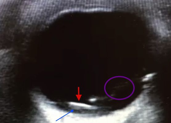
Point of care ultrasound revealed a mobile, radiolucent hyperechoic structure (see red arrow) with reverberation within the posterior chamber (see blue arrow), likely a metallic foreign body. Linear areas of mobile hyperechoic material revealed possible vitreous hemorrhage (see purple circular area). Orbital non-contrast CT confirmed a 3 mm metallic focus within the dependent portion of the left globe, lodged in the posterior sclera, with some vitreous hemorrhage but no evidence of globe rupture. Ophthalmology was consulted and the patient was taken to surgery later that night.
OphthalmologyVisual EM
Creative Commons images

Computed Tomography Diagnosis of Appendicitis

Christopher Libby, MPH* and Shannon Toohey, MD, MAEd^

DOI: https://doi.org/10.21980/J8F30NIssue 2:1[mrp_rating_result]
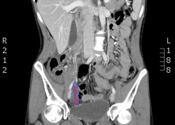
The CT abdomen/pelvis with IV contrast shows a dilated appendix (see red outline) with thickened, hyperenhancing wall (see blue outline) best visualized in the axial and coronal planes.
Abdominal/GastroenterologyVisual EM
Creative Commons images

Intracranial Hemorrhage Following a 3-week Headache

John Jiao, MHS* and Alisa Wray, MD*

DOI: https://doi.org/10.21980/J89885 Issue 2:1[mrp_rating_result]
The patient’s head CT showed a significant area of hyperdensity consistent with an intracranial hemorrhage located within the left frontal parietal lobe (red arrow). Additionally, there is rightward midline shift up to 1.1cm (green arrow) and entrapment of the right lateral ventricle (blue arrow).
NeurologyVisual EM
Creative Commons images

Gastric Bezoar

Samer Assaf MD*

DOI: https://doi.org/10.21980/J85K5WIssue 2:1[mrp_rating_result]
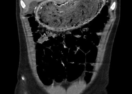
In the abdominal radiograph, a nonspecific and non-obstructive bowel gas pattern with no air-fluid level was noted, however the stomach was distended with soft tissue. The CT abdomen/pelvis revealed a distended stomach with undigested heterogeneous contents (presumed bezoar).
Abdominal/GastroenterologyVisual EM
Creative Commons images

Monteggia Fracture in an Assault Patient

John Jiao, MHS* and Shannon Toohey, MD, MAEd*

DOI: https://doi.org/10.21980/J81S3ZIssue 2:1[mrp_rating_result]
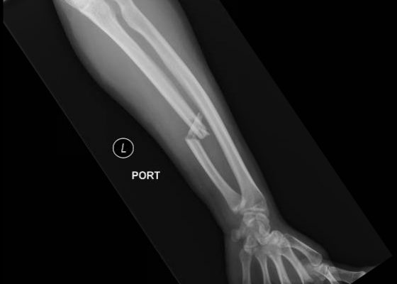
On the axial elbow x-ray, the radial head (red arrow) is dislocated anteriorly from the humerus; the humeroulnar articulation is intact. On the AP forearm x-ray, there is a closed, displaced, comminuted fracture of the ulna (blue arrow).
OrthopedicsVisual EM
«‹79›»
Page 79 of 91

JETem is an online, open access, peer-reviewed, journal-repository for EM educators. We are PMC Indexed.

Most Viewed

  • Simple Auricular Hematoma (“Cauliflower Ear”) Task Trainer
  • The Three Breath Method: Teaching Coping Mechanisms for Processing Patient Death
  • Clinical Decision Making: Acute Ischemic Stroke with Large Vessel Occlusion
  • PRIMER: Premedical Introduction to Mentored Emergency Research: Connecting Undergraduate Assistants and Emergency Medicine Residents in a Community Hospital Setting
  • Dangerous Dysrhythmias in the ED: An Interactive EKG Workshop for Emergency Medicine Clerkship Learners

Visit Our Collaborators


Creative Commons Licence
This work is licensed under a Creative Commons Attribution 4.0 International License.

About

Education

Learners should benefit from active learning. JETem accepts submissions of team-based learning, small group learning, simulation, podcasts, lectures, innovations, curricula, question sets, and visualEM.

Scholarship

We believe educators should advance through the scholarship of their educational work. JETem gives educators the opportunity to publish scholarly academic work so that it may be widely distributed, thereby increasing the significance of their results.

Links

  • Home
  • Aim and Scope
  • Current Issue
  • For Reviewers
  • Instructions for Authors
  • Contact Us

Newsletter

Sign up to receive updates from JETem regarding newly published issues and findings.

Copyright Creative Commons Attribution 4.0 International