
Latest Articles

Meeting the Milestones through Simulation: Disclosing a Medical Error
DOI: https://doi.org/10.5070/M5.53010By the end of this simulation activity, learners will be able to: 1) disclose a medical error to a patient and/or their family, 2) understand the different types of patient safety events that should be reported, 3) locate the form for disclosing a patient safety event at their institution, and 4) appropriately complete a patient safety report at their institution.
Simulation-Based Interprofessional Didactic for Emergency Management of Snakebites
DOI: https://doi.org/10.5070/M5.61957By the end of the simulation, participants will be able to recognize and appropriately manage neurotoxic snakebite envenomation, including initiating first-line management and toxicology consultation, coordinating antivenom administration, recognizing and treating antivenom-associated anaphylaxis, assessing and managing progressive respiratory failure, demonstrating effective interprofessional communication using TeamSTEPPS principles,4 and coordinating timely consultation, handoff, and disposition with toxicology, pharmacy, and the critical care team.

How to Place a Central Line: Preparing Your Kit & Performing the Procedure
DOI: https://doi.org/10.5070/M5.54074By the end of these videos, learners will be able to:
1. Identify and gather all necessary materials for internal jugular (IJ) CVC placement.
2. Describe the appropriate positioning of the patient, ultrasound machine, and proceduralist for CVC placement.
3. List common complications of IJ CVC placement.
4. Demonstrate sterile field preparation, including donning personal protective equipment (PPE), placing a sterile drape, and placement of a sterile ultrasound probe cover.
5. Perform a proper pre-procedural time-out including at least two patient identifiers and site verification.
6. Prepare and organize CVC kit components for efficient procedural flow.
7. Identify key anatomical landmarks and optimal patient positioning for right IJ CVC placement using ultrasound guidance.
8. Demonstrate stepwise needle insertion with real-time ultrasound visualization to safely cannulate the
internal jugular vein.
9. Verify proper guidewire placement using both short- and long-axis ultrasound views before vessel dilation.
10. Perform safe dilation and catheter advancement using standard procedural technique.
11. Exhibit best-practice behaviors, including port flushing, line securement, and sterile dressing application.
12. Document the procedure and identify post-placement safety steps such as confirmatory chest imaging.

Transforming Emergency Medicine Mentorship: A Skill-Building Curriculum to Optimize Mentee Growth and Development
DOI: https://doi.org/10.5070/M5.52383The aim of this curriculum is to train mentors to approach mentoring with a curious and non-judgmental mindset. The curriculum offers specific, practical exercises to increase mentors’ ability to foster a mentee-driven relationship that meets the needs of all mentees, which in turn enhances career development and faculty retention at the department level.

Ruptured Hemorrhagic Ovarian Cyst Mimicking Ruptured Ectopic Pregnancy in a Post-Transplant Patient: A Case Report
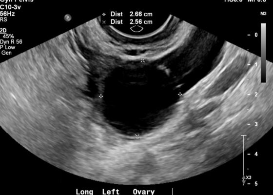
DOI: https://doi.org/10.5070/M5.52390A formal transvaginal ultrasound was ordered in the setting of the positive beta-hCG levels, which demonstrated a moderate volume of debris-containing free pelvic fluid layering in the posterior cul-de-sac (yellow highlighting), without sonographic evidence of an intrauterine gestational sac or yolk sac. Additionally, the left ovary contained a 2.8 cm cystic structure consistent with a hemorrhagic ovarian cyst (blue highlighting).
Intentional Pencil Ingestion – A Case Report of a Sharp Ingested Foreign Body
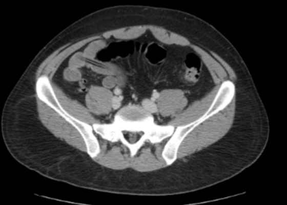
DOI: https://doi.org/10.5070/M5.53005Computerized tomography (CT) of the abdomen and pelvis two days after the ingestion of the pencil revealed a hypodense foreign object measuring 7.9 cm (blue arrows) in the lumen of the distal ileum. The images obtained demonstrate that the object progressed past physiological strictures within the gastrointestinal tract such as the esophagus and stomach; however, it had yet to progress through the ileocecal junction. Ingested foreign bodies carry a high risk of obstruction and perforation at these physiological stricture points. A repeat CT scan six days later showed that the pencil (yellow arrow) had progressed to the distal transverse colon.
Case Report: Sonographic Detection of Chorionic Hemangiomas in Sturge-Weber Syndrome
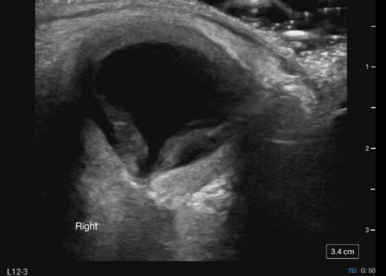
DOI: https://doi.org/10.5070/M5.52314Ocular point-of-care ultrasound (POCUS) was performed using a linear 12-3 MHz probe on a Sonosite PX system. Examination of the symptomatic right eye revealed a complete retinal detachment. The detached retina appeared thickened, consistent with chronic detachment. Deep to the retina, the choroid was markedly thick with the widest portion observed in the posterior wall of the globe near the optic nerve. Sonographically, the choroid was isoechoic to the retina in the near field, hyperechoic to the retina in the far field (secondary to posterior acoustic enhancement) and homogenous. In the colorized image of the right eye, the red color denotes the choroid, labeled C, and the green color denotes the retina, labeled R. The left eye was also examined, shown in the last case image, and was grossly normal. No color doppler imaging was utilized for either eye. Optic nerve sheath diameter (ONSD) was 0.54 cm bilaterally.
Point-of-Care Ultrasound Findings of Ovarian Hyperstimulation Syndrome: A Case Report
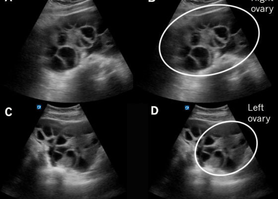
DOI: https://doi.org/10.5070/M5.52324Transabdominal POCUS in the right upper quadrant revealed clearly delineated pockets, the hypoechoic appearance being consistent with ascites, surrounding the liver and the gallbladder (Figure 1). A view of the left upper quadrant revealed a similar hypoechoic fluid collection that filled the space below the diaphragm and surrounded the spleen (Figure 2). A transabdominal pelvic view revealed enlarged ovaries, identifiable by the numerous, large, and well-delineated hypoechoic collections within that represent the enlarged follicles (Figure 3). The extent of the hypoechoic free fluid collections and the enlargement of the ovaries and their follicles is more apparent when viewed dynamically (Video).
Case Report: Atypical Presentation of Large Bowel Obstruction Due to Bezoar Formation in a Patient with a History of Colon Cancer
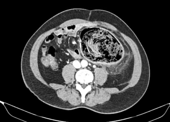
DOI: https://doi.org/10.5070/M5.53997CT with intravenous (IV) contrast revealed: (1) a large amount of fecal material in the descending colon at the level of anastomosis suggesting bezoar formation, (2) colonic wall thickening and inflammation suggestive of stercoral colitis, (3) abrupt caliber change at distal descending colon indicating partial large bowel obstruction, and (4) moderate to severe left hydronephrosis due to ureteropelvic junction obstruction with 0.4 cm calculus. No abscess or pneumatosis visualized. Annotated images demonstrate a large intraluminal mass at the descending colon anastomosis with associated abrupt caliber change (arrow), causing partial LBO and adjacent colonic wall inflammation (highlighted) consistent with stercoral colitis.
Case Report on Rare Presentation of Intravenous Leiomyomas with Intracardiac Leiomyomatosis
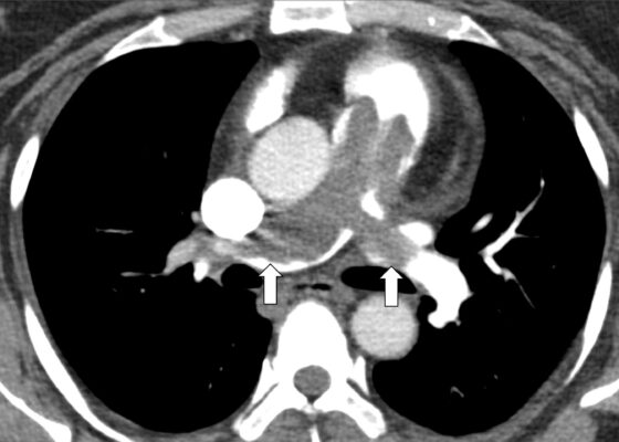
DOI: https://doi.org/10.5070/M5.52111Postoperatively, a CT of the abdomen and pelvis with contrast showed a lobulated soft tissue density (arrowheads) measuring 4.5 x 3 cm in the right pelvis. A subsequent MRI of the pelvis showed a 3.7 x 2.8 cm lobulated structure in the right pelvis above the vaginal cuff thought to be most consistent with an unusual cluster of venous structures. Following this, a CT venogram of the abdomen and pelvis revealed residual filling defects in the infrarenal IVC, extending through the right common iliac vein and multiple internal iliac branches.