
JETem
  • Home
  • About
    • Aim and Scope
    • Our Team
    • Editorial Board
    • FAQ
  • Issues
    • Current Issue
    • Ahead of Print
    • Past Issues
  • Visual EM
    • Latest Visual EM
    • Search Visual EM
    • Thumbnail Library
  • For Authors
    • Instructions for Authors
    • Submit to JETem
    • Photo Consent
    • Policies
      • Peer Review Policy
      • Copyright Policy
      • Editorial Policy, Ethics and Responsibilities
      • Conflicts of Interest & Informed Consent
      • Open Access Policy
      • Acceptance Fee
  • For Reviewers
    • Instructions for JETem Reviewers
    • Interested in Being a JETem Reviewer?
  • Topic
    • Abdominal/Gastroenterology
    • Administration
    • Board Review
    • Cardiology/Vascular
    • Certifying Exam Cases
      • Clinical Decision-Making
      • Prioritization
      • Communication
      • Procedure & Ultrasound
    • Clinical Informatics, Telehealth and Technology
    • Dermatology
    • EMS
    • Endocrine
    • ENT
    • Faculty Development
    • Genitourinary
    • Geriatrics
    • Hematology/Oncology
    • Infectious Disease
    • Miscellaneous (stats, etc)
    • Neurology
    • Ob/Gyn
    • Ophthalmology
    • Orthopedics
    • Pediatrics
    • Pharmacology
    • Procedures
    • Psychiatry
    • Renal/Electrolytes/Nutrition
    • Respiratory
    • Social Determinants of Health
    • Toxicology
    • Trauma
    • Ultrasound
    • Urology
    • Wellness
    • Wilderness
  • Modality
    • Certifying Exam Practice Cases
      • Clinical Care Cases
      • Communication & Procedural Cases
      • Old School Oral Board Cases
    • Curricula
    • Innovations
    • Lectures
    • Podcasts
    • Simulation
    • Small Group Learning
    • Team Based Learning (TBL)
    • Visual EM
  • Contact Us

Visual EM

Creative Commons images

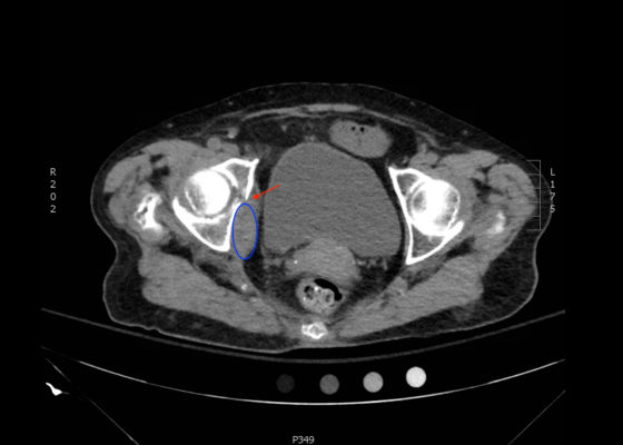
Acetabular Fracture

Chad Correa* and Sari Lahham, MD^

DOI: https://doi.org/10.21980/J8BK8K Issue 2:4[mrp_rating_result]
The non-contrast CT images show a minimally displaced comminuted fracture of the right acetabulum involving the acetabular roof, medial and anterior walls (red arrows), with associated obturator muscle hematoma (blue oval).
OrthopedicsVisual EM
Creative Commons images

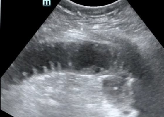
Bedside Ultrasound for the Diagnosis of Small Bowel Obstruction

Alexander Anshus, BS* and Maili Alvarado, MD^

DOI: https://doi.org/10.21980/J86W6PIssue 2:4[mrp_rating_result]
The POCUS utilizing the low frequency curvilinear probe demonstrates fluid-filled, dilated bowel loops greater than 2.5cm with to-and-fro peristalsis, and thickened bowel walls greater than 3mm, concerning for SBO. 
Abdominal/GastroenterologyVisual EM

Chancre of Primary Syphilis

Adnan Riaz MD* and Grant Wei MD*

DOI: https://doi.org/10.21980/J83342 Issue 2:4[mrp_rating_result]
Physical examination revealed a non-tender, erythematous lesion on the glans penis, two similar adjacent satellite lesions, as well as tender inguinal lymphadenopathy. No penile discharge was noted.
Infectious DiseaseDermatologyGenitourinaryVisual EM
Creative Commons images

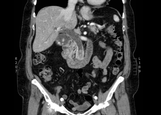
Choledocholithiasis

Jonathan Peña, MD*

DOI: https://doi.org/10.21980/J8Q62X Issue 2:3[mrp_rating_result]
Computed tomography (CT) was significant for two large gallstones measuring 1.1 centimeters impacted at the level of the pancreatic head with associated common bile duct (CBD) dilatation.
Abdominal/GastroenterologyVisual EM
Creative Commons images

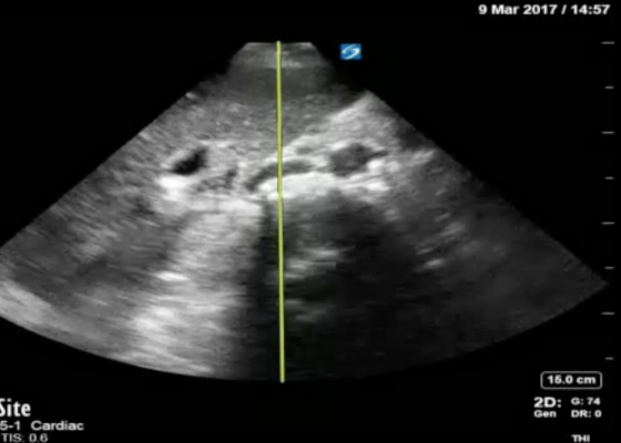
Using Bedside Ultrasound to Rapidly Differentiate Shock

Mohamad Moussa, MD*, Jacob Willen, BS* and Sudipa Biswas, BS, MS*

DOI: https://doi.org/10.21980/J8S047 Issue 2:3[mrp_rating_result]
A RUSH exam demonstrated hyperdynamic cardiac contractility and collapse of the inferior vena cava (IVC) with probe compression more than 50% suggesting hypovolemia likely secondary to sepsis. Incidentally, Morrison’s pouch revealed a large right renal cyst but no signs of free fluid. A computed tomography of abdomen/pelvis showed a 10.8 x 9.5 cm right renal cyst and left lower lobe pneumonia.
UltrasoundInfectious DiseaseVisual EM
Creative Commons images

Corneal Sparing Conjunctival Abrasion

Hamid Ehsani-Nia, MS*, Tanya Dall, MD^ and Samantha Costantini, MD^

DOI: https://doi.org/10.21980/J8KG93 Issue 2:3[mrp_rating_result]
Physical exam was significant for multiple broken teeth, multiple minor abrasions on the face, and fine shards of shattered glass on his face and hair. His right eye had conjunctival injection, with no signs of subconjunctival hemorrhage or ocular penetration. Vision, extraocular movement, and pupillary exam were grossly intact. Fluorescein staining with slit lamp exam with cobalt blue filter examination of the right eye revealed superficial bulbar conjunctival uptake of fluorescein dye staining an area of the conjunctiva inferior to the limbus 5 mm vertical by 2 mm horizontal (estimation by photo provided). No foreign bodies were visualized in the inferior fornix. These findings were consistent with superficial conjunctival abrasion. The exam noted sparing of the corneal epithelium.
OphthalmologyVisual EM
Creative Commons images

Volvulus

Sari Lahham, MD, MBA*, Kathryn Bennett, BS* and Mohammad Helmy, MD†

DOI: https://doi.org/10.21980/J8JH0Q Issue 2:3[mrp_rating_result]
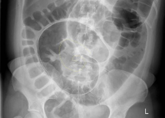
Upright and supine frontal radiographs of the abdomen demonstrate gas dilation of the large bowel from the level of the cecum to the sigmoid colon with air fluid levels (yellow arrows). There is a swirled configuration of the distal descending to sigmoid colon indicating the level of the volvulus (dashed yellow line) and giving rise to the classic “coffee bean” sign (dotted white tracing). Note the elevated left hemidiaphragm on the upright view reflecting abdominal distention with increased intra-abdominal pressure (red arrow).
Abdominal/GastroenterologyVisual EM
Creative Commons images

Pneumonia Diagnosed by Point-of-Care Ultrasound

Akash Patel, BA*, Steven Bunch, MD^, Maili Alvarado, MD^, Shadi Lahham, MD^ and J. Christian Fox, MD^

DOI: https://doi.org/10.21980/J8XP64 Issue 2:3[mrp_rating_result]
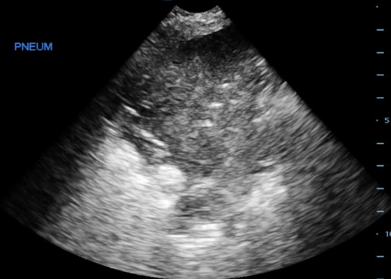
Point-of-care ultrasound of the left lower lobe demonstrates lung hepatization, a classic finding for pneumonia. In addition, a shred sign is present with both air bronchograms and focal B-lines—all suggestive of poorly aerated, consolidated lung. The patient was started on antibiotics and admitted to the hospital with a diagnosis of community-acquired pneumonia.
UltrasoundRespiratoryVisual EM
«‹37›»
Page 37 of 47

JETem is an online, open access, peer-reviewed, journal-repository for EM educators. We are PMC Indexed.

Most Viewed

  • Simple Auricular Hematoma (“Cauliflower Ear”) Task Trainer
  • The Three Breath Method: Teaching Coping Mechanisms for Processing Patient Death
  • Clinical Decision Making: Acute Ischemic Stroke with Large Vessel Occlusion
  • PRIMER: Premedical Introduction to Mentored Emergency Research: Connecting Undergraduate Assistants and Emergency Medicine Residents in a Community Hospital Setting
  • Dangerous Dysrhythmias in the ED: An Interactive EKG Workshop for Emergency Medicine Clerkship Learners

Visit Our Collaborators


Creative Commons Licence
This work is licensed under a Creative Commons Attribution 4.0 International License.

About

Education

Learners should benefit from active learning. JETem accepts submissions of team-based learning, small group learning, simulation, podcasts, lectures, innovations, curricula, question sets, and visualEM.

Scholarship

We believe educators should advance through the scholarship of their educational work. JETem gives educators the opportunity to publish scholarly academic work so that it may be widely distributed, thereby increasing the significance of their results.

Links

  • Home
  • Aim and Scope
  • Current Issue
  • For Reviewers
  • Instructions for Authors
  • Contact Us

Newsletter

Sign up to receive updates from JETem regarding newly published issues and findings.

Copyright Creative Commons Attribution 4.0 International