
Visual EM
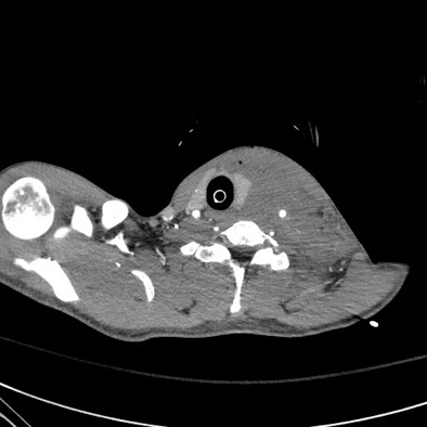

A Case Report of a Transected Carotid Artery Caused by a Stab Wound to the Neck
DOI: https://doi.org/10.21980/J8BP8MThe post intubation chest x-ray (CXR) showed severe rightward displacement of the trachea (purple arrow). The computed tomography angiogram (CTA) showed transection of the left common carotid artery (LCCA), extensive neck hematoma without extravasation and severe tracheal deviation to the right (blue arrow). The intravenous (IV) contrasted chest computed tomography (CT) image showed a lateral contrast projection from the aortic arch at the level of the isthmus (green and pink arrows). There were no other significant injuries reported on the CT scans of the chest, abdomen and pelvis.

Case Report of COVID-19 Positive Male with Late-Onset Full Body Maculopapular Rash
DOI: https://doi.org/10.21980/J86W72The images demonstrate a diffuse, flat, maculopapular exanthema along the torso, bilateral upper and lower extremities, and neck without edema consistent with reported cutaneous manifestations of COVID-19. There are no surrounding bullae, vesicles, or draining. On palpation, there was blanching of the rash. Sensation to light touch was intact in all extremities. The findings were also apparent on the face with no mucosal involvement.

Posterior Sternoclavicular Dislocation: A Case Report
DOI: https://doi.org/10.21980/J8363QChest X-ray revealed an inferiorly displaced right clavicle at the right sternoclavicular joint (blue arrow). A computed tomography angiogram (CTA) of the chest was therefore obtained and revealed a right posterior sternoclavicular dislocation with resultant compression of the left brachiocephalic vein (purple arrow). Even though the right clavicle is displaced, the anatomy of the brachiocephalic vein is such that it is positioned to the right of midline, placing the left brachiocephalic vein posterior to the right clavicle. The right brachiocephalic and common carotid artery were normal in appearance. The CTA also revealed a comminuted fracture of the left anterior second rib at the costochondral junction that had not been previously seen on the x-ray.
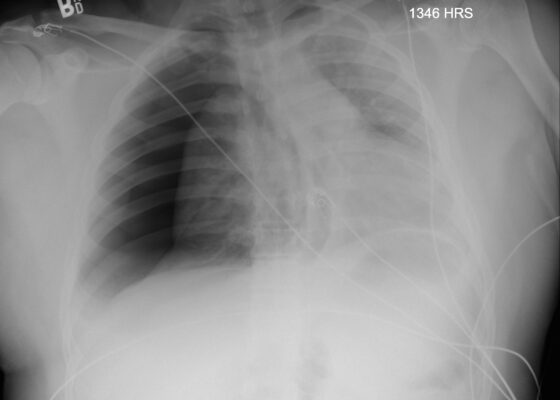
Case Report: Traumatic Tension Pneumothorax in a Pediatric Patient
DOI: https://doi.org/10.21980/J8ZD1SChest X-ray demonstrated significant right-sided pneumothorax (with red outline showing border of collapsed right lung) with cardio mediastinal shift to the left (shown by blue arrows) indicative of a tension pneumothorax
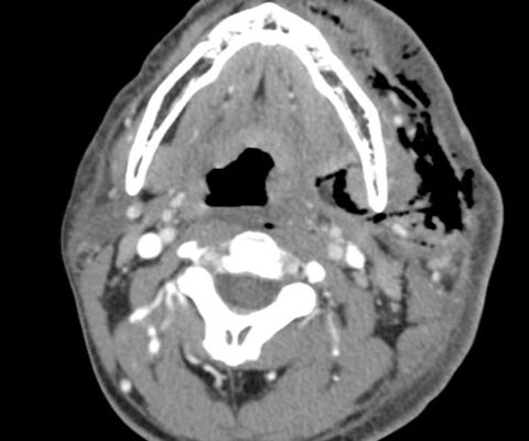
Necrotizing Fasciitis and Mediastinitis after Wisdom Tooth Extraction: A Case Report
DOI: https://doi.org/10.21980/J8XW7KComputer tomography (CT) imaging of soft tissues of the neck and of the chest/abdomen/pelvis revealed extensive swelling and subcutaneous air (see red arrows) on the left side of the face and neck extending to the left shoulder, as well as parapharyngeal/retropharyngeal spaces and posterior/superior mediastinum.

Adult Clavicular Fracture Case Report
DOI: https://doi.org/10.21980/J8FM0TThe patient's chest and clavicular radiographs showed a comminuted displaced acute fracture of the right mid-clavicle (green, blue, yellow). The clavicular fracture was also visible on the chest computed tomography (CT). The remainder of his trauma workup was negative for acute findings.

Case Report of Distal Radioulnar Joint and Posterior Elbow Dislocation
DOI: https://doi.org/10.21980/J89S6KRadiographs of the left elbow and wrist were obtained. Left elbow radiographs showed simple posterolateral dislocation of the olecranon (red) without fracture of the olecranon (red) or trochlea (blue). Left wrist lateral radiographs demonstrated DRUJ dislocation with dorsal displacement of the distal ulna (green) without fracture or widening of the radioulnar joint (purple). Post-reduction radiographs demonstrated appropriate alignment of the elbow with the trochlea seated in the olecranon and improved alignment of the DRUJ.
Bladder Diverticulum – A Case Report
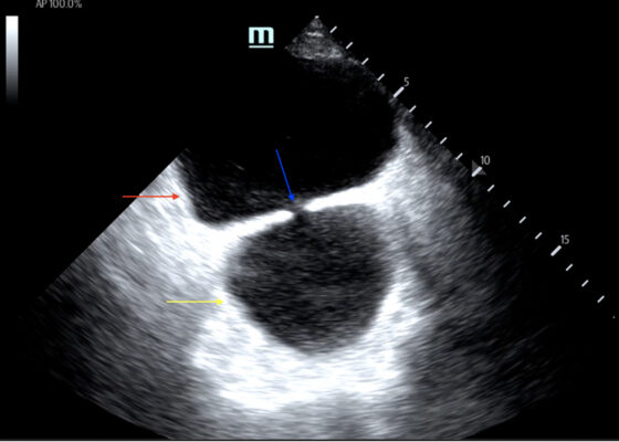
DOI: https://doi.org/10.21980/J8635COn examination, the patient was alert and oriented but in mild distress. Suprapubic fullness was noted upon abdominal palpation. Point of care ultrasound of the bladder showed two enlarged “bladders” with a central communication. Bedside total bladder volume was measured to be 1288 cm3 (the top “bladder” was measured to be 1011 cm3, while the bottom “diverticulum” was measured to be 277 cm3) by ultrasound.
The POCUS stills of the patient’s bladder demonstrated the bladder (red arrow) and bladder diverticulum (yellow arrow) with a central communication (blue arrow) in the transverse and sagittal views.