Ophthalmology
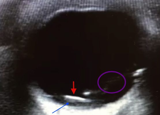

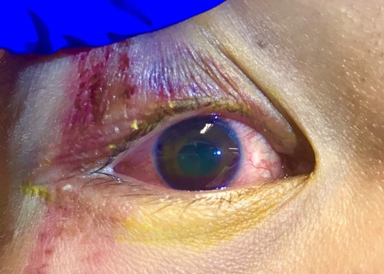
Intraocular Foreign Body: Ultrasound and CT Findings
DOI: https://doi.org/10.21980/J8JS3MPoint of care ultrasound revealed a mobile, radiolucent hyperechoic structure (see red arrow) with reverberation within the posterior chamber (see blue arrow), likely a metallic foreign body. Linear areas of mobile hyperechoic material revealed possible vitreous hemorrhage (see purple circular area). Orbital non-contrast CT confirmed a 3 mm metallic focus within the dependent portion of the left globe, lodged in the posterior sclera, with some vitreous hemorrhage but no evidence of globe rupture. Ophthalmology was consulted and the patient was taken to surgery later that night.

The Acute Red Eye
DOI: https://doi.org/10.21980/J8BC74By the end of this educational session, the learner will: 1) list 10 major causes for an acute red eye; 2) describe historical features that help distinguish between benign and serious causes of the acute red eye; 3) describe physical examination features that help distinguish between benign and serious causes of the acute red eye; and 4) use historical and physical examination features to distinguish between the 10 different causes of the acute red eye.